Bluebell: Hyacinthoides non-scripta

Standard

[This post features my original artwork and botanical sketches]

I truly believe that revising for my upcoming exams and developing my understanding of the sciences is an enjoyable process, but when gentle dawn sunlight illuminates the vibrant azure bundles of flowers peppered throughout our garden, it takes the strength of every fibre in my being to not abandon my textbooks and immerse myself in the carpet of wild bluebells.

In two months, I’ll have sat all my AS exams and will finally be free to laze around in the grass with my cat and lie among the sea of indigo petals. Until then, I look to the shimmering haze of the bluebells as an oasis just fingertips away.

bluebell - Edited (3) (1)

Recently, I rewarded my revision efforts by pressing an inflorescence and uncovering the chemistry behind their beautiful blue pigment*. The rich colour of the petals is not caused by a single true blue pigment but rather by the red pigment anthocyanin. The basic structure of the anthocyanin delphinidin is modified with pH shifts and prosthtic groups.

bluebell - Edited (4) (1)Delphinidin is pH sensitive and turns red in acidic environments. A high pH must be maintained to ensure the molecular pigment is blue. Delphinidin is also malonated; two glucosyl groups, a malonyl group, and a coumaryl group are bonded to the pigment structure. 


The resulting compound, malonylawobanin, is responsible for the brilliant deep blue petals that don’t even fade after weeks of pressing. 

I also consulted my copy of The Complete Herbal out of interest in the medicinal properties of this plant. Nicholas Culpeper was surprisingly brief in his writings of bluebells; he only refers to its somewhat styptic (blood clotting) qualities. From further research, I learnt that the bluebell’s intensely toxic nature has, understandably, limited its potential for therapeutic applications.

One of the major toxic compounds present in bluebells are polyhydroxylated pyrrolidines (such as nectrisine) which are found in the viscous sap that seeps out of the plant’s nodding stems. These compounds are analogues of sugar but are actually potent glucosidase inhibitors. Due to its many -OH groups, the mammalian body mistakes nectrisine as a sugar and processes it as such – inadvertantly allowing the molecule to interfere with respiration.

In addition to this, polyhydroxylated pyrrolizidines, which have a similar chemical action to nectrisine, are toxic alkaloids that have been proven to poison livestock that have the misfortune of ingesting bluebells. Despite this, these compounds are now being considered for use in cancer therapeutics.

Most fascinatingly, bluebells contain a type of cardiac glycoside, Scillaren A, which can rapidly increase the output force of the heart. Heart cells, myocytes, contain protein pumps embedded in their cell membranes which actively transport sodium ions out of the cell to create an essential electrochemical gradient. In the heart, there is also a sodium – calcium ion exchanger which maintains the ion homeostasis. 

Cardiac glycosides, such as Scillaren A found in bluebells, inhibit the movement of sodium ions out of the cell by effectively disabling the ion pumps. This increases the concentration of sodium ions inside the myocytes which, in turn, increases the calcium ion concentration as sodium ions inside the cell are exchanged for calcium ions.

In myocytes, the accumulation of calcium increases the power with which the cells can contract (contractility) – increasing cardiac muscle contraction which can be crucial in controlling the heart rate in cases of arrhythmia and even chemically restarting the heart in atrial fibrillation.

The cardiac action of Scillaren A is stronger than that of the commonly used digoxin, which is derived from foxglove, and is ideal for situations in which digoxin alone is insufficient or the patient has a digitalis intolerance. Additionally, if an improper dosage is administered, Scillaren A is poisonous but, unlike many other cardiac glucosides, is non-lethal as it is poorly absorbed into the gut. The compound also has a very high therapeutic index as it is rapidly eliminated from the body. All of these qualities make bluebells a fundamentally desirable pharmaceutical source, however, since native bluebells are an endangered and protected species, bluebells cannot be harvested to produce medication.

bee - Edited (1)

Conservation efforts are essential to protect and preserve this fragile, flowering species so that we may be able to enjoy their magnificent appearance and benefit from their therapeutic value in the coming decades.


* White bluebells which lack pigmentation also exist albeit they are somewhat rare with only 1 in every 1000 native bluebells being white. Below is a pressed white bluebell I came across in Margate. bluebell - Edited (2) (1)

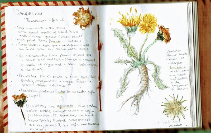
Dandelion: Taraxacum Officinale

Standard

[This posts features my original botanical sketches]

For me, the most marvellous manifestations of spring are the bright dandelions emerging from previously barren, frost-glazed soil – bringing promises of brighter days to come.  The humble dandelion is so often maligned as a weed, however these perennial plants have a rich therapeutic history.

dandelion - Edited (2) (1)

Nearly half a millennium ago, Nicolas Culpeper, a prominent herbalist of the early 1600s, noted that the dandelion has “an opening and cleansing quality” and remarked upon its efficacy at treating liver ailments (including cirrhosis) as well as diseases of the spleen and gall-bladder. Many of Culpeper’s contemporaries, and indeed modern herbalists, also acknowledge the powerful diuretic properties of dandelion leaves and root.

Dandelions have even proven valuable in cutting-edge pharmaceuticals. Researchers from the Department of Chemistry and Biochemistry at the University of Windsor, Canada,  have found that dandelion root extract can efficiently kill certain types of aggressive leukaemia cells. This extract can induce apoptosis (programmed cell death) and autophagy (wherein the cancer cell breaks down its own organelles), and even disrupt mitochondrial membranes. The lack of toxins and alkaloids in the plant, combined with the extract’s selectivity for leukaemia cells, makes the dandelion a particularly promising candidate for safe chemotherapy applications.


Ovadje, P., Hamm, C., & Pandey, S. (2012). Efficient induction of extrinsic cell death by dandelion root extract in human chronic myelomonocytic leukemia (CMML) cells. PloS one7(2), e30604. doi:10.1371/journal.pone.0030604

https://www.ncbi.nlm.nih.gov/pmc/articles/PMC3281857/


The Shape Of Water

Standard

Water is the most abundant molecule in cells, organism, and on earth and it is so vital to life due to a combination of its unique chemical and physical properties.

Let’s first take a closer look at the structure of this essential molecule; water is polar (meaning that it has small charges on the individual atoms in the molecule which let it bond to other water molecules). This is so important and influences all other properties of water. Because water is polar, it can interact with other substances (such as salt) to dissolve them. In our bodies, all the chemicals are dissolved in water in our cells (in the form of the cytoplasm): water allows us to exist at a cellular level.


hb

The solid form of water (ice) demonstrates another cool property of water as ice is actually less dense than its liquid form. We probably all take this surprising physical property of water for granted, however, for all other substances, the opposite is true. Ice is less dense than water because when the liquid cools down and molecules lose energy, more hydrogen bonds form between the molecules- forcing the molecules into a more spread out lattice to accommodate for the maximum amount of hydrogen bonds.

VPf

This unique physical property ensures that, even when its really cold, bodies of water such as lakes and oceans freeze from top-down – insulating the water below the surface and protecting aquatic life. This special property of water might have prevent organisms from going extinct in the past.

Water is so important to life that it is no wonder that we are almost constantly told to remain hydrated, especially in the summer. Naturally, in order to transport water, the majority of us use plastic water bottles: they’re convenient, lightweight, and cheap. But their hidden cost comes in the form of the damage it does to our health and the health of the planet.

The fact that plastic poses a risk to the environment probably doesn’t come as a shock to you. Despite this, you would probably still be forced to use plastic bottles since there are virtually no other cheap, lightweight alternatives that can even hold a candle to plastic. Until now…

Ooho

Introducing Ooho! A water bottle that is completely edible and 100% biodegradable. Developed by Skipping Rocks Lab and inspired by the natural membranes found in eggshells, these orbs of water are encased in a calcium alginate gel layer and formed through a fascinating process known as spherification which was developed, not by chemists, but by chefs.

The process of spherification was pioneered in the 1950s by Unilever and it involves the reaction of two common chemicals which can form a tough membrane on the outside of the water- essentially, the water became its own water bottle through polymerisation. The first ingredient of the edible ‘water bottle’ is Sodium Alginate, usually derived from seaweed. This is a long-chain carbohydrate that is soluble in water. In its original plant, it is used to store sugars created by photosynthesis. When Sodium Alginate is dissolved in water, these long-chain carbohydrates float around on their own but they don’t connect to each other. That’s because poking out from these long chains is a branch of carbon and oxygen atoms, which chemists call an anionic group which has a slight negative charge. The sodium ions are attracted to this, because they have a positive charge. But sodium is monovalent, meaning that it wants to bond to just one of these carbon and oxygen branches at a time.

However, if you add calcium ions to the solution, the structure changes. Calcium ions have two positive charges, so they want to bond onto two of these branches at a time, meaning that it can connect two alginate molecules together. So the calcium ions replace the sodium and create cross-linkages between the long-chain carbohydrates. When enough of these chains are connected together, this 3D matrix of connected alginate chains forms a semi-solid impermeable membrane that the water can’t pass through -creating a sphere that surrounds the water. The fully formed water sphere is made from nothing but water and the two chemicals!

spherificationpt1-1editeditver2

membrane

As part of the session, we experimented with sodium alginate and calcium lactate to design our own edible ‘water bottles’. We first dissolved one gram of sodium alginate powder into 250 ml of water to create sodium alginate solution, Then we dissolved five grams of calcium lactate powder into 1250 ml of water to create a calcium lactate bath. We then gently set a tablespoon of the sodium alginate solution into the calcium lactate bath- allowing the spoon to become completely immersed before tipping it over to pour out the sodium alginate solution.

The main difficulty was estimating when to remove the water orbs from the calcium lactate bath; we came to the conclusion that 20 minutes is the minimum amount of time needed for the membrane to solidify properly whereas leaving the ‘water bottles’ submerged for an hour ensured that the membrane remained flexible but strong.

Avenues for further investigation would be to freeze the sodium alginate into spheres and then submerge the solid spheres into the calcium lactate bath to produce ‘water bottles’ with a more defined shape and to experiment with calcium chloride which is said to produce a stronger membrane. Overall, however, I feel the experiment was fairly successful and we were able to sample numerous ‘water bottles’!

Capture

20181112_151047

This technique is causing a bit of a stir in manufacturing circles as it could provide a waste-free way to contain and transport water as the membrane is both edible and biodegradable. It is still a long way from replacing the water bottle on store shelves, however, as the gel membrane breaks down over time and isn’t as tough as plastic.


This blog post is based on the course content I created for a series of sessions on the biochemistry of water as part of the Exploring Everyday (Bio)Chemistry Society which I run at The Tiffin Girls’ School. The sessions focus on connecting chemistry with everyday products and phenomena to encourage KS3 engagement with science and they have been a great success so far. More blog posts based on this society can be found here.

A Toast to the Maillard Reaction

Standard

Earlier this year, I wrote an article on the Maillard Reaction and the science that underpins toast and ageing for the Oxford Scientist Schools Writing Competition in response to the prompt “How does science impact your everyday life?”.

This morning, I was delighted to find that my article was selected as a runner-up and published on the Oxford Scientist website! You can read it here or below…


We all have our little morning rituals. I, for one, greet the day by performing a series of complex chemical reactions – and so do you. I am, of course, referring to  the Maillard Reaction which takes place in practically every kitchen and café throughout the country. We have this beautiful cascade of reactions to thank for the rich, invigorating aroma of freshly roasted coffee and the golden glow of toast.

The Maillard reaction refers to a chain of reactions between amino acids and reducing sugars and it is named after the prominent French chemist Louis-Camille Maillard who first investigated these reactions in 1912.  Subsequent chemists, such as John E Hodge in 1973, further researched the culinary significance of these reactions and their impact on  flavour, aroma, and appearance.

When a slice of bread first enters the confines of a toaster, the temperature of the heating coils quickly rise to an optimum 154° C  at which point the system is provided with enough thermal energy to begin the Maillard reaction. The reactive carbonyl group on a reducing sugar reacts with the amino group of an amino acid within the bread to produce a nitrogen-substituted glycosylamine molecule.  The glycosylamine molecule then isomerises to produce a ketosamine which undergoes further reactions depending on the environment.

flat,800x800,070,f.u5

The slightly acidic conditions of white bread (which typically has a pH of 5.0 – 6.2) allows for the formation of melanoidins. Melanoidins are long, polymeric pigments (much like the melanin in our skin). In more alkaline conditions, the melanoidins are produced faster and in a greater quantity – resulting in a deeper shade of brown.

Recipes for Bavarian-style pretzels manipulate the Maillard reaction to create the glossy brown glaze of the baked goods; the dough is first dipped in weak lye solution to increase the alkalinity of the coating before baking.

During the Maillard reaction, hundreds of other organic compounds may be formed depending on a variety of factors such as the types of amino acid present, and the temperature and pH of the environment. When coffee is roasted, heterocyclic aromatic compounds are formed- including furans (which give coffee its caramel-like bittersweet scent) and pyrazines (which add earthy notes to coffee’s flavour profile). Due to its sheer complexity, the Maillard reaction can result in an impressive range of potential flavours – to the delight of chefs and chemists alike.

Whilst you were musing over the complex science responsible for enhancing the flavours of your breakfast, if you had completely forgotten about the bread in the toaster, you would become acquainted with the darker side of the Maillard reaction. As the temperature rises and the bread is subjected to high levels of thermal energy for a longer period of time, a potentially carcinogenic product, acrylamide, is formed. Such  compounds are a natural consequence of cooking, however as the bread suffers for even longer within the toaster, reactions such as pyrolysis become more prevalent and the concentration of acrylamide in the toast increases- leaving behind an acrid taste in your mouth.

Surprisingly, the Maillard reaction impacts our lives even after we have left the kitchen; at a much slower rate, it is constantly occurring within our bodies – forming a range of potentially deadly, undesirable products: advanced glycation end products (AGEs).  Over the course of a lifetime, the accumulation of AGEs on DNA can lead to neurodegeneration and degenerative eye diseases (including cataract formation). Researchers are currently looking into preventing ocular degeneration and reducing the effects of ageing through inhibition of the Maillard  reaction.

The Jekyll and Hyde nature of the Maillard reaction has  a profound impact on our everyday lives. It gives us the gift of golden toast, the manna of students everywhere; however, in our golden years, the very same reaction accelerates our inevitable decline. Currently, the formation of insidious products during the Maillard reaction is unavoidable. Instead of fearing ageing and burnt toast, we should strive for a deeper knowledge of the intricacies of the Maillard reaction which will allow us to work with its innate chemistry- making our everyday lives healthier and more delicious.

4055-3-large

In The Lab: Synthesising Polymers

Standard

In one of the earlier sessions of Exploring Everyday (Bio)Chemistry Society, we discussed the properties of natural polymers (including spider silk). Our focus now moves on to artificial polymers (including rayon and nylon). During our experiment based sessions  of this term, we replicated one of the earliest methods of synthesising rayon (a semi-synthetic fibre designed 125 years ago by the English chemist, Charles Frederick Cross).

First, a solution of tetra-amine-copper(II) ions must be prepared; I measured out 10 grams of basic copper carbonate and, working under a fume hood, added 100 cm3 of 880 ammonia solution. Despite operating in one of the best-ventilated labs, the choking scent of ammonia wrapped its tendrils around my airways almost immediately- suffocating me. I can safely say that the outcome of this experiment was still definitely worth it!

IMG_20190114_085919IMG_20190114_090808


[Carefully measuring out the copper carbonate and dissolving it in ammonia to form a solution of tetra-amine-copper stirr.gifions]

Next, I added 1.5 grams of finely shredded cotton wool to the solution. The cotton wool provides the cellulose (a natural reactant) for the reaction- which is why rayon is only a semi-synthetic polymer.


When cotton is dissolved in this solution, the resulting mixture becomes surprisingly viscous (with an almost shower gel-like consistency).

This initially presented a slight problem in achieving complete dissolution- a problem which was solved with the help of an exciting device I’d never had the chance to use before: a magnetic stirrer.

A magnetic stirrer consists of a small bar magnet which is dropped into the solution and a device which creates a rotating magnetic field – forcing the bar magnet to rotate and, hence, stir the solution.

The resulting effect is that the solution appears to stir itself – much to the shock and delight of some of the younger attendees of the session!


The final step is to simply inject a thin stream of the deep blue solution under the surface of some sulfuric acid.  As the solution enters the acid, its pH drops swiftly and the complex compound is converted into insoluble rayon.

ezgif.com-video-to-gif (1)


Although the rayon initially appears deep blue in colour, as the copper (II) ions diffuse out of the rayon (reacting with the sulfuric acid to form copper sulfate solution), the colour quickly fades away- leaving behind only the delicate, translucent fibres.

IMG_20190114_130439              IMG_20190114_142217


Unraveling the Secrets of Spider Silk – 1

Standard

Until quite recently, I’ve always considered myself as a bit of an arachnophobe – the very thought of a spider’s unnerving eight-legged dance across the floor is enough to send a tsunami of shivers down my spine.

In the past few months, however- I’ve been forced to interact with spiders (and their handiwork) more than I would have necessarily wanted to. Last November, my cat greeted me with sunlight glinting off the gossamer wisps trailing from her whiskers, purring contently having just devoured another of her newfound culinary delights – spider webs. Although superficially endearing and quirky, the risk of her tucking in to a web coated with the toxin 2-pyrrolidinone (a form of chemical warfare some spiders wield against predatory ants and other creatures that threaten their webs) had compelled me to assess the toxicity of the webs that have accumulated in neglected corners of the garden.

In general, threatening spiders such as the black widow and brown recluse spin unconventional web formations such as the tangle web – infamous for its haphazard arrangement of strong silk threads. Examining the structure of the spider webs my cat gravitates to every morning has allowed me, for the first time, to truly appreciate the intricacies of arachnid architecture. As I retreated back to my office, to a more comfortable distance from the nearest spider, a wave of security washed over me and, intrigued, I started to research the synthesis and spinning of spider silk.

Spider silk is a natural polypeptide which is technically classed as a scleroprotein (a broad category of structural proteins which encompasses collagen (which is found in our ligaments) and keratin (in our nails and hair). There are many different types of spider silk which are chemically designed to suit its purpose; dragline silk, for instance, is used to create the radiating support lines of the web structure and is also used to connect the spider to the web. Swathing silk is a special silk used to wrap and immobilise the spider’s prey. Spiders even create a unique silk for ‘parachuting’ which is released into the air and caught by the wind- allowing the spider to fly!

To enable them to produce so many different types of silk, spiders have a surprisingly complex anatomy; the underside of their abdomen has three to four separate silk glands containing a watery fluid of amino acids known as ‘dope’. Microscopic tubes pump the dope to spinnerets (appendages which solidify and spin the silk). The silk leaves the spider through spigots – protrusions with muscular valves – which control the diameter of the silk fibres. The faster and tighter the strand is drawn, the stronger the silk.

spigot

Spigots emitting silk (Gasteracantha sp.), SEM, [Credit: Dennis Kunkel]

Whilst the exact chemical composition of these proteins depends on a combination of genetic and environmental factors, including the species of spider and its diet, the main protein in spider silk is fibroin which consists predominantly of two amino acids: glycine and alanine. The elasticity of spider silk is due to glycine-rich regions of the protein chain which allow the protein to fold in on itself to form a beta-spiral structure.  Capture silk, the most elastic kind of spider silk, has a very high concentration of glycine and so can extend up to 4 times its original length.


Because of its fantastic properties, it is no surprise that scientists want to harvest spider silk and combine it with other materials to design strong, lightweight bulletproof vests and very flexible, biodegradable rope. The issue is that, unlike silkworms, spiders cannot be farmed to produce large quantities of silk as they are cannibalistic and would resort to eating their neighbours if they were forced into close proximity of each other. Additionally, spider silk is so fine that more than 400 spiders would be needed just to produce a square metre of the material.

Scientists have therefore been trying to recreate spider silk in the lab; chemical synthesis of spider silk is not yet viable since we still do not fully understand the silk structure. Currently, replication of silk is achieved through genetic engineering- silk genes have been inserted into Escherichia coli bacteria to successfully produce the proteins spidroin 1 and spidroin 2. But an unforeseen flaw cropped up; the silk produced by the bacteria was surprisingly weak due to faulty cross-linking  in the protein structure.

The next avenue of research is inserting the silk genes into goats to produce spider silk proteins in their milk since milk production in mammary glands is similar to silk protein production in spiders so proper protein cross-linking could be achieved in goats.


Keep Calm and Curry On

Standard

If you’ve ever had the pleasure of experimenting in the kitchen and making curries, you are probably already familiar with turmeric – you might even have a jar of the golden spice hiding in your kitchen cupboards right now. Although not usually the first substance to come to mind when thinking about chemistry, turmeric (and its active agent curcumin) have some truly fascinating chemical properties. 

As with all spices, turmeric is a mix of chemical compounds, but its instantly recognisable bright yellow colour is due to the presence of a particular compound: curcumin. 

curcuminCurcumin is a polyphenol (an organic compound with multiple phenol units).  The phenol units and aromatic structures of curcurim mean that the electrons in the curcumin molecules can absorb (and, hence, gain energy from) UV light. The energy gained promotes them to a higher energy level (an ‘excited state’). This is the same excitation that occurs during flame tests of metal ions. This process doesn’t last long though; they quickly lose some of this energy as vibrational energy, before dropping back down to their original energy level, emitting their excess energy as visible light. This means that curcumin can fluoresce!

In order to demonstrate this amazing ability of curcumin, I swirled turmeric into a beaker of ethanol (as the spice is more soluble in organic solvents) and viewed the solution under a blacklight- the results were almost mesmerising.

ezgif.com-video-to-gif.gif

Another exciting property of curcumin is its ability to act as a chemical pH indicator (like universal indicator solution or the natural compound anthocyanin). Curcumin changes colour in alkali solutions because of a subtle change in its chemical structure.

Curcumin’s chemical structure contains a sequence of alternating single and double bonds between atoms, what chemists refer to as ‘conjugation’. The length of this sequence of alternating bond types affects the wavelengths of light that the compound absorbs. In curcumin’s case, when it’s added to an alkaline solution, a subtle change in structure alters the length of the conjugated part of the molecule, in turn altering the wavelengths of light that it absorbs. This causes the colour change from yellow to red.

capture


I also demonstrated this property by adding turmeric to a beaker of dilute HCl and a beaker of dilute KOH. In a more informal (kitchen) setting, this experiment could also be replicated with lemon juice (which is weakly acidic) and bleach.

img-20181105-wa0002

Finally, much to the delight of the attendees, we created pH indicator paper from the turmeric-ethanol solution and filter paper; the homemade indicator paper works within seconds, quickly providing answers to some of life’s greatest mysteries such as what is the pH of coffee, seawater, and even saliva!


Thank you very much to Compound Interest for the brilliant article and presentation which this session of Exploring Everyday Bio(Chemistry) was based on.

Check out their video (with more details of the experiment and great animations) below.


This blog post is based on the course content I created for a series of sessions on the chemistry of spices as part of the Exploring Everyday (Bio)Chemistry Society which I run at The Tiffin Girls’ School. The sessions focus on connecting chemistry with everyday products and phenomena to encourage KS3 engagement with science and they have been a great success so far. More blog posts based on this society can be found here.


Wake Up And Smell The Solvents

Standard

A thick blanket of petroleum ether fumes had descended upon the lab- interspersed with the acrid scent were traces of delicate citrus notes. This was the surprising result of my attempt to extract essential oil from oranges via expression and solvent extraction.  Despite producing an essential oil that didn’t quite resemble the uplifting citrusy scent of oranges, I left the lab with a smile that stretched for miles – ecstatic about having designed and carried out a complete experiment entirely single-handedly as part of an exciting club I have recently set up at school.

The idea of organising a club had been slowly growing in my mind since the start of the summer holidays. Having just finished all my GCSE exams, it was almost expected that I would want to completely avoid any curricular activities; however, I remained passionately interested in the subjects I chose to study at Sixth Form and started really looking forward to studying A-Level Chemistry in particular. Curious as to what the A-Level course would be like, I came across the FutureLearn course on ‘Exploring Everyday Chemistry’ run by the University of York. In six weeks, I was exposed to the fascinating organic chemistry and biochemistry that underpinned so many commonplace products and concepts- ranging from fragrances to beverages.


My mind started brimming with terms such as ‘limonene’, ‘enantiomers’, and ‘vibrational bond energy’. I decided on modifying the content of the course to start up our school’s very own Exploring Everyday Chemistry Society; the concept of connecting chemistry with everyday products is a brilliant one that would encourage engagement with science (particularly for younger pupils in KS3 and 4).

What makes this proposed club unique is that, each half-term, I planned on carrying out and leading experiments to enhance the course content and help younger students (and myself) develop vital practical skills.

Which brings us to this afternoon; I had the wonderful experience of demonstrating the extraction limonene essential oil from orange peel to Year 9 student.


img-20181008-wa0015

First, working over a 250 cm3 beaker, we used the grater to remove only the outer, coloured rind from the oranges and scraped the material from the grater into the beaker with a spatula.

Next, the gratings were gathered into the garlic press and squeezed into a conical flask through a strainer.


img-20181008-wa0012

20 cm3 of distilled water was then added to the flask and the liquid was transferred to a separating funnel. We then carefully added 10 cm3 of solvent (petroleum ether) and shook the separating funnel before leaving the emulsion to separate out into two layers.

The bottom layer (mostly water and debris) was run off and discarded and the top layer (consisting of petroleum ether and dissolved essential oil) was transferred into a water bath to evaporate off the petroleum.


img-20181008-wa0010

Due to time limitations and using excess petroleum ether, the final collected essential oil still contained a slight amount of solvent.

I did find that allowing the liquid to evaporate at room temperature reduced the intensity of the petrol vapours. However, when I repeat this experiment with the Year 10 students next week- I’ll ensure to use less solvent and boil off the solvent thoroughly.


Overall, this experiment is a great success to me and has been an experience I enjoyed tremendously – we produced 16 little vials of essential oil to take home and treasure.

(I still wouldn’t use them as perfume though…)

20181008_150325