A Sixth Sense – the science behind Migraines with Aura

Standard

So Much More Than ‘Just A Headache’

Unfamiliar with the extreme pain a migraine brings, it’s natural to try and relate it to a bad headache – mistaking the condition’s throbbing pain for a common distracting ache. Besides intense pain, migraines also come with a host of sensory issues such as severe sensitivity to sound and light (known as photophobia), neck stiffness, and visual phenomena. While headaches are an infamous symptom of illness, migraines are the illness.

The most striking difference between a migraine and a headache is that, almost as if they have a sixth sense, one in three migraine sufferers can predict when one is brewing before the pain sets in. Relax – we’re not omniscient. In the build up to a migraine attack, we experience auras: transient visual disturbances that are as disorientating as they are fascinating from a scientific perspective.

What starts out as a shimmering haze in the migraine sufferer’s periphery, gradually envelops their field of vision with flashing lights, shimmering arcs, and blind spots. Although auras serve as a warning for an impending migraine attack, they can be far more dangerous than the ensuing headache; during auras, brain cells can become damaged, even to the point of death, causing ischaemic strokes and creating lesions in the white matter of the brain. But what’s the connection between a migraine and these strange visual symptoms?

Origin of an Aura

At the onset of a migraine, the biochemistry of the brain changes drastically. At rest, healthy neurons contain fewer positively charged ions such as sodium ions (Na+) and potassium ions (K+) and more negatively charged proteins and nucleic acids than their surroundings. This separation of charge gives their membranes a low resting potential of -70 mV.
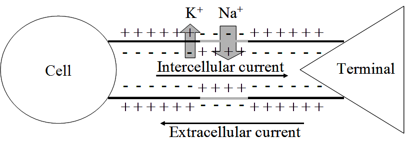
If we compare a neuron’s axon to a simple tube connecting one neuron to its neighbour, we can see that the inside of the axon is more negative than its surroundings outside the cell (the extracellular space).

Triggering the migraine aura is a sudden, spontaneous efflux of K+ ions out of a single neuron buried deep in the brain’s visual cortex – causing the cell’s membrane potential to plummet. The build-up of K+ ions in the extracellular space triggers the release of amino acid glutamate which excites neighbouring neurons and allows K+ ions to flood in – propagating the ion movement further. On the heels of this sudden flurry of activity, comes a wave of neuronal silence in which each cell stops processing and transferring information as it slowly returns to normal.
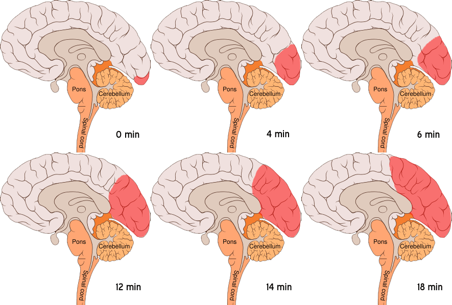
This wave of intense excitement followed by inactivity spreads across the visual cortex – corrupting visual information from the eyes and creating strange auras.

This phenomenon, known as cortical spreading depression (CSD), is responsible for the peculiar auras that slowly take over a migraineur’s visual field. When a neuron is affected by the spreading depression, it stops accurately processing visual information from the eyes – instead interpreting signals into disturbances like flashing lights or blurry blind spots.

The gradual spread of CSD across the visual cortex is mirrored by the gradual growth of an aura in the build up to a migraine attack.

Cortical spreading depression is not unique to the visual cortex; it can occur throughout the grey matter. The reason why this wave of electrical activity originates in the visual cortex is because this region of the brain is very densely crammed with neurons – increasing the risk of one neuron’s aberrant activity affecting the many cells around it.

CSD & Pain

The effects of CSD extend far beyond interfering with vision. Neuron excitation in the visual cortex activates adjacent trigeminal nerves which span your face from temple to jaw. These nerves run parallel to major blood vessels of the meninges (the cushioning tissue between your skull and brain) and, when activated, trigger the release of pain-generating substances into the bloodstream. As a result, the meninges become inflamed, causing blood vessels to widen and put further pressure on the tissue – intensifying the pain.

To make matters worse, these waves of neural activity up-regulate the expression of genes encoding pro-inflammatory molecules (such as COX-2, the target of many painkillers) exacerbating the sensation of pain. Additionally, CSD activates metalloproteinases, provoking the blood-brain barrier to leak; pro-inflammatory molecules, as well as the K+ and glutamate from earlier, are now free to flood into the major facial blood vessels. In combination, these molecules sensitise the nerves in the neck and jaw, causing the notorious aches and stiffness many migraine sufferers endure. This sensitisation is also why normally innocuous processes (like coughing and yawning) hurt and cause tension to build up during a migraine.
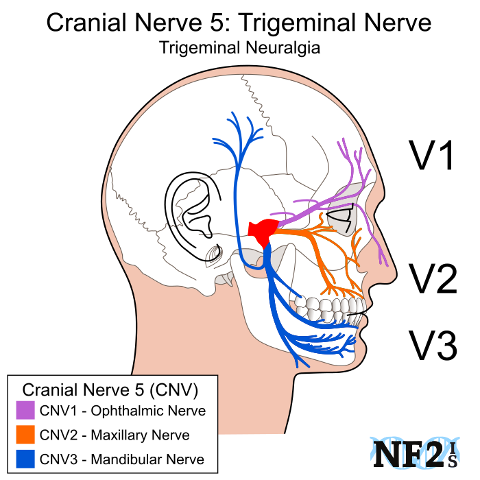
The spreading depression is first transmitted to the ophthalmic nerve branch and is then propagated to the mandibular nerves, bringing the pain along with it.

Potential Treatments

None of these excruciating effects are felt in ordinary headaches where CSD plays no role; to reiterate, the intense pain felt by migraine sufferers comes down to the unique biology of a migraine attack – it has nothing to do with low pain thresholds and its far more than just a bad headache.

Currently, the majority of migraine treatments either suppress pro-inflammatory molecule production or tackle the excessive blood vessel widening which puts pressure on the meninges. While they are useful in dealing with the headache aspect of a migraine attack, they have no effect on auras and fail to address the root cause of the migraine: the CSD.

Drugs that interfere with dopamine and serotonin pathways have been proven effective, quite possibly because they control neurotransmitter release and hence limit excitation of adjacent neurons in CSD – curbing the spread of depolarisation. Potential novel treatments also address the K+ ion channel dysfunction that triggers CSD, preventing both the aura and the pain that follows it.
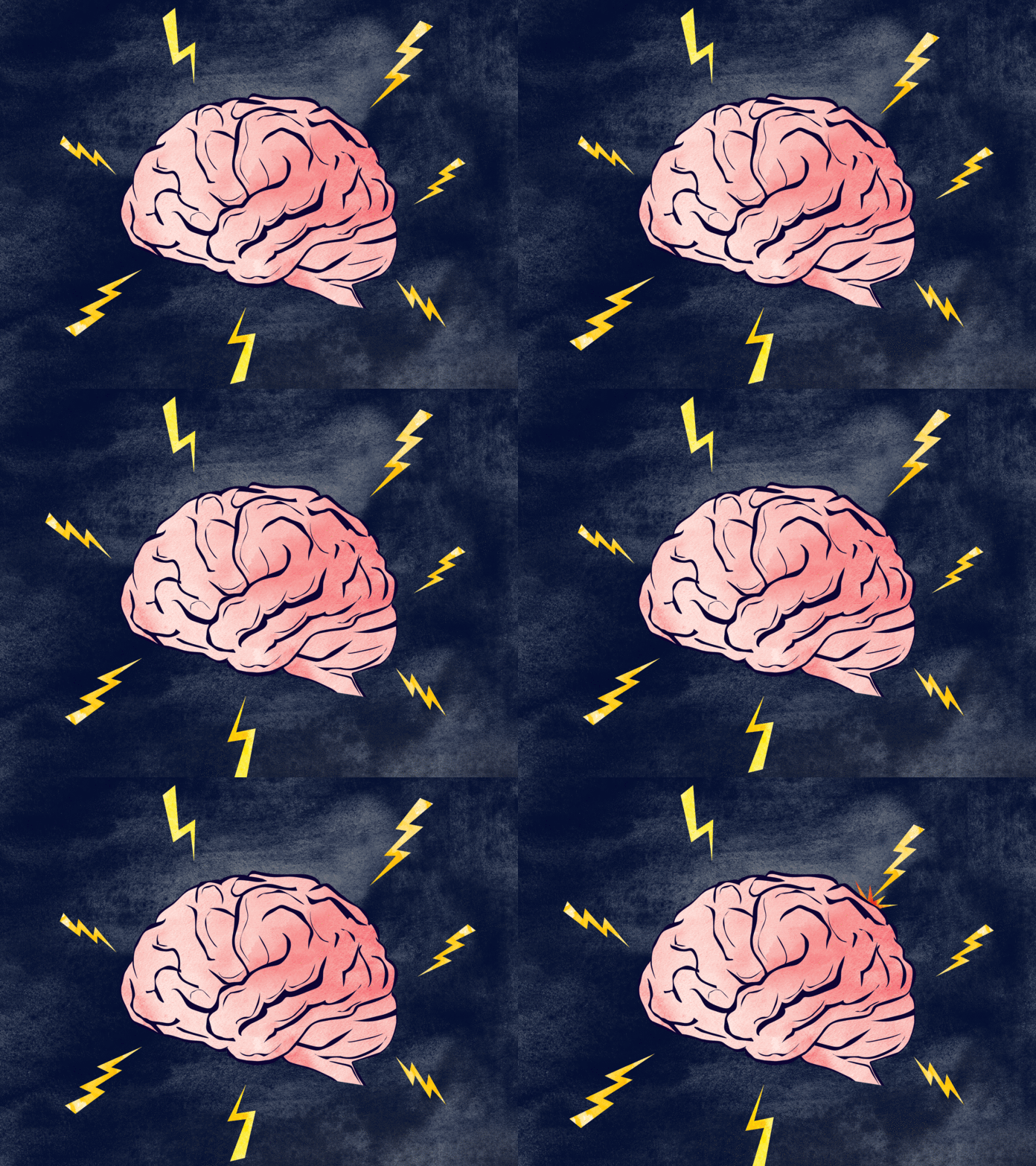
When it comes to migraine treatment, the classic proverb rings true – prevention is better than cure. However, there are shockingly few effective migraine prophylactics.
Research into what triggers the K+ ion efflux of CSD in the first place and why sufferers are genetically predisposed to having migraines could spare the nearly 10 million of us in the UK from frequent bouts of agony.

.

Bibliography:

  • Pietrobon, D., Striessnig, J. Neurobiology of migraine. Nat Rev Neurosci 4, 386–398 (2003). https://doi.org/10.1038/nrn1102
  • Lauritzen, M. (1987). Cortical spreading depression as a putative migraine mechanism. Trends in Neurosciences, 10(1), 8–13. doi:10.1016/0166-2236(87)90115-9 
  • Charles, A. C., & Baca, S. M. (2013). Cortical spreading depression and migraine. Nature Reviews Neurology, 9(11), 637–644. doi:10.1038/nrneurol.2013.192 
  • Medscape.com. (2019). Migraine Headache: Practice Essentials, Background, Pathophysiology. [online] Available at: https://emedicine.medscape.com/article/1142556-overview#a3.
  • Spierings, E. L. H. (2001). MECHANISM OF MIGRAINE AND ACTION OF ANTIMIGRAINE MEDICATIONS*. Medical Clinics of North America, 85(4), 943–958. doi:10.1016/s0025-7125(05)70352-7 

I’ve written this post for Migraine Awareness Week – if you wish to support research into treatments for this debilitating neurological condition, consider donating to Brain Research UK.

The Chemistry of Communication

Image

Action_potential_propagation_animation

This brilliant gif, created by John Schmidt, summarises how ions cause changes in voltage that allow electrical signals to be propagated down axons. The accompanying explanation of the gif clarifies the many factors at play here:

Three types of ion channel are shown: potassium “leak” channels (blue), voltage-gate sodium channels (red) and voltage-gated potassium channels (green). The movement of positively-charged sodium and potassium ions through these ion channels controls the membrane potential of the axon.

The negative-inside resting potential is mostly determined by potassium ions leaving the cell through leak channels. Action potentials are initiated in the axon initial segment after a neurotransmitter activates excitatory receptors in the neuron’s dendrites and cell body.

This depolarizes the axon initial segment to the threshold voltage for opening of voltage-gated sodium channels. Sodium ions entering through the sodium channels shift the membrane potential to positive-inside, approaching the sodium equilibrium potential.

The positive-inside voltage during the action potential in the initial segment causes the adjacent part of the axon membrane to reach threshold. When positive-inside membrane potentials are reached, voltage-gated potassium channels open and voltage-gate sodium channels close.

Potassium ions leaving the axon through voltage-gated potassium channels return the membrane potential to negative-inside values near the potassium equilibrium potential. When the voltage-gated potassium channels gate shut, the membrane potential returns to the resting potential.

(Credit: Wikiversity)


This post  is part of the Neuroscience Crash Course (a mini series about the brain created in preparation for the Brain Bee) and is a further clarification of A Closer Look at Neurons – I would strongly recommend reading that post before this one.

A Closer Look at Neurons

Standard

The previous post explained the nervous system- a circuit which transfers messages throughout the body. Now it is time to look even more closely at the nervous system and, in particular, then the individual units of its circuitry- the neurons.

The neuron is the basic working unit of the brain. The specialized cell’s function is to transmit information to other nerve cells, muscle, or gland cells. The human brain contains approximately 100 billion neurons!

All neurons consist of a cell body, dendrites, and an axon.  The axon extends from the cell body and ends in several nerve terminals. Dendrites also extend from the neuron cell body and receive messages from other neurons.

378156c3cf901f8868bac4c86ee238ecd434d9bb

Basic structure of a neuron (Credit: Bitesize)


The point of contact between two neurons is called a synapse- dendrites are covered with synapses where they connect with the nerve terminals of other axons.

eJpbb

(Credit: Biology Stack Exchange)

In order to send messages to other neurons, electrical impulses are transmitted along the axons. Just like in a computer, the efficiency of electrical transmissions  in the brain is imperative. In order to ensure this, axons are covered with a layered myelin sheath that helps accelerate transmissions. (If you are interested in how the myelin sheath can do this, I’ve explain this in further detail at the end of the post.) Glia (a type of specialised cell) form the myelin sheath. The naming of glia varies depending on where they are found; in the brain, they are called oligodendrocytes, and in the peripheral nervous system, they are known as Schwann cells.

glia-

(Credit: Lumen Learning)

Glia are essential for the proper functioning of the brain as they perform many important roles such as transporting nutrients to neurons, cleaning up brain debris, digesting parts of dead neurons, and helping hold neurons in place. Surprisingly, there are ten times more glia than neurons in the brain.

credit-quora

A microscopy image to put things into perspective (Credit: Quora)


Communication in the brain requires nerve impulses which involve the opening and closing of ion channels. Ion channels are molecular, water-filled tunnels which pass through the cell membranes of neurons and allow ions (which are electrically charged) to enter or leave the cell. As ions flow through an ion channel, an electric current is created- resulting in small differences in voltage across the neuron’s cell membrane.

This difference in charge is essential for allowing a neuron to generate an electrical impulse. When a nerve impulse is initiated, the neuron switches from a negative charge state to a positive charge state (causing a “dramatic reversal” in the electrical potential of the cell). This change, known as action potential, then passes along the axon’s membrane. The action potential can travel at impressive speeds of up to hundreds of miles per hour which allows neurons to generate electrical impulses multiple times every second.

action-potential-crash-course

(Credit: Crash Course)

When the action potential reaches the end of an axon, it triggers the release of neurotransmitters. Neurotransmitters released at nerve terminals diffuse across the synapse and bind to receptors on the surface of the target cell (i.e. neuron or  a muscle or gland cell). This interaction alters the electrical potential of the target cell’s membrane and triggers a response from the target cell. The response can range from the generation of an action potential to the contraction of a muscle.

Neurotransmitters are an important area of research as they could help us understand even more of the brain. Learning about the various chemical circuits of the brain could help explain how memories are created and stored as well as what is responsible for disorders such as Alzheimer’s and Parkinson’s diseases.

The next post will be all about this fascinating chemical messenger.


The myelin sheath is essential for speeding up transmission. Instead of merely propagating continuously down the axon, the action potential is generated at the bare regions (known as the nodes of Ranvier) where there is no myelin (found between myelinated segments of the axon).


myelin2

(Credit: hyperphysics.phy-astr.gsu.edu)


This post  is part of the Neuroscience Crash Course (a mini series about the brain created in preparation for the Brain Bee) .

Sending Signals: How the brain relays information

Standard

The first post of this crash course explored the areas of the brain and their functions. This post will delve deeper into how areas of the brain communicate with each other and the rest of the body.

The spinal cord (an extension of the brain which is about 17 inches long and protected by the vertebral column) receives sensory information and connects the brain to the rest of the body below the head. The sensory information it receives is used for reflex responses to pain as well as generating nerve impulses in nerves that control the muscles and organs through voluntary commands from the cerebrum.

The nervous system is a network of nerve cells and fibres which transmits nerve impulses between parts of the body. If you crave a more eloquent definition, in Brain Facts the nervous system is defined as

“a vast biological computing device formed by a network of gray matter regions interconnected by white matter tracts” – Louis Vera-Portocarrero

There are two great divisions of the nervous system: the central nervous system (CNS) and the peripheral nervous system (PNS). The CNS consists of the forebrain, midbrain, hindbrain, and spinal cord. The PNS consists of nerves and small concentrations of grey matter called ganglia.

PNSCNS

The PNS can be further divided into the somatic nervous system and the autonomic nervous system.

The somatic nervous system is comprised of neurons which connect the CNS with the parts of the body that interact with its surroundings (the sensory organs and voluntary muscles). Somatic nerves in the cervical region control the neck and arms; those in the thoracic region serve the chest; and those in the lumbar and sacral regions interact with the legs.

regions

Somatic nerve regions (Credit: Brain Facts)

The autonomic nervous system is comprised of neurons connecting the CNS with internal organs and it is also divided into two parts: the sympathetic nervous system (which provides energy and resources during times of stress and arousal) and the parasympathetic nervous system (which conserves energy and resources during relaxed states).

nervoussystem

This diagram explains the nervous system and its many subdivisions very clearly (Credit:Psychology Hacked)


Messages are carried throughout the nervous system by neurons. The next post will explore the structure of neurons and how they transmit and receive messages.


This post  is part of the Neuroscience Crash Course (a mini series about the brain created in preparation for the Brain Bee) .

A Tour of the Brain

Standard

The human brain, despite its vast complexities, has a relatively straightforward architecture. This post will explore the structure of one of the most important organs of the body.

Imagine that you are looking at a human brain sitting on the lab worktop before you.

At first sight, the brain appears to consist entirely of a large mass of spongy tissue. These deeply folded outer layers of the brain make up the cerebral cortex and take up nearly two thirds of the entire volume of the brain. Observe the seemingly erratic pattern of curved  grooves on the surface (the sulci) and you will be able to identify the major divisions of the cerebral cortex. Divided into two hemispheres (which are bridged by a bundle of fibres called the corpus callosum), the cerebral cortex houses the majority of the ‘grey matter’ and is separated into a few important lobes.

The first lobes you notice are the frontal lobes. They are aptly named considering that they are found at the front of the brain (near the forehead). The lobes (one in each hemisphere) are responsible for decision making, planning, memory, voluntary action, and even personality.

fl

(Credit: Wikipedia)

Next, you spot the parietal lobes which are located at the crown of the head. These lobes are heavily involved in perception and interpretation of all sensory information. They are also necessary for spatial awareness and attention.

pl

(Credit: Wikipedia)

Below the parietal lobes lie the occipital lobes. Found at the back of the brain, the occipital lobes are mainly involved in vision. Damage to the occipital lobes often leads to blindness as well as other vision-related defects.

The last of the main divisions of the cerebral cortex are the temporal lobes. In vivo, they are found near the ears. The temporal lobes are essential for object recognition, memory formation, and language.

tl

(Credit: Wikipedia)

Now imagine picking up the brain (whilst wearing latex gloves- naturally). Raise it above eye level and you will see the prominent brain stem emerging from the base of the brain. In vivo, this would be connected to the spinal cord- linking the brain to the body.

Nestled just behind the brain stem is the cerebellum. Literally meaning ‘little brain’, the cauliflower-shaped brain structure plays an important role in not only movement, fine motor control, and posture but also in memory, mood, and language processing as well as Pavlovian learning. The cerebellum is comprised of two hemispheres which are connected via a narrow structure known as the vermis.

ins.JPG

(Credit: Quizlet)

Now place the brain back down on the worktop and carefully pry apart the hemispheres to observe the hidden inner structures of the brain. Remember the brain stem? Look above it and you will discover the midbrain and then, above the midbrain, the egg-shaped thalamus. Heavily interconnected with several other regions in the cortex, most sensory information first passes through here before moving on to the relevant parts of the cortex.

Our last stop on this tour of the brain is the hippocampus. To access this, you must cut into the temporal lobes as this seahorse shaped region is located deep within these lobes. The hippocampus is highly involved in the formation and consolidation of memories and in spatial navigation. Damage to the hippocampus can cause severe amnesia.

hip

(Credit: Pinterest)

You have now observed all of the basic architecture of the brain. Knowing this makes it much easier to comprehend diseases of the brain and to discover cures.


This post  is part of the Neuroscience Crash Course (a mini series about the brain created in preparation for the Brain Bee).

Next post in the Neuroscience Crash Course