Cats Are Not Peas : Review & Research

Standard

Cats Are Not Peas is a popular science book regarding the history of genetics and how the field evolved from early theories of fertilisation and trait inheritance to a modern understanding of gene expression and disorders. The common theme running through the book is the author, Laura Gould’s, quest to better understand her rare male calico cat, George.   

Ginger cats are generally male and calico (aka. tortoiseshell) cats are almost never male. The allele for a ginger coat colour is recessive and the coat colour gene is carried on the X chromosome. Female cats have two X chromosomes, so, in order for a female to be ginger, she must have the ginger allele on both of her X chromosomes- which is unlikely in a typical feline gene pool. If she has only one  ginger allele, she will usually be a tortoiseshell cat. At the sixty-four-cell stage of embryonic development, a form of dosage compensation known as X-inactivation randomly silences one X chromosome of the pair in each embryonic cell, independently of neighbouring cells. The silenced chromosome is maintained as a Barr body for all future cell divisions. So each cell has either an active ginger allele or an active black allele – resulting in a coat that is a mosaic of the two coat colours. 

Since Cats Are Not Peas is written for a non-specialist audience, Gould does not delve into the details of Barr body formation. However, this piqued my interest and I researched X-inactivation further. Calico cats were actually involved in the history of X-inactivation study; in 1949, Murray Barr and his PhD student Ewart Bertram identified a dark, condensed structure (a Barr body) close to the nucleolus in the dissected motoneurons of a female calico cat. In 1961, Mary Lyon formulated her X-inactivation theory which proposed a process which leads to the global silencing of the entire X chromosome. This prompted further investigation into how embryonic cells can ‘count’ the number of X chromosomes to inactivate (always equal to (n-1) chromosomes). For instance, in an XX pair only one chromosome is silenced whereas in the case of triple X syndrome two X chromosomes are silenced. The blocking hypothesis suggests that ‘counting’ is achieved by small amounts of a blocking factor binding to the Xic locus of a single X chromosome in the pair. The other X chromosome(s) will then be inactivated as they are unblocked. 

Another line of investigation regarded the ‘choice’ of which chromosome to inactivate; what were the molecular mechanisms of skewed choice or randomness? Non-random choice can be a result of secondary selection for/against certain embryonic cells – if an allele on the active chromosome leads to cell death, all surviving cells in the embryo will have that X chromosome inactivated. Alternatively, primary skewing of the X inactivation choice may be a result of certain X chromosomes being more resistant to inactivation. A key example of this is the X-controlling element (Xce) found in mice. In 1972, Bruce Cattanach identified that resistance to X-inactivation is influenced by which Xce allele each chromosome had.

Returning to the book, calico cats are not simply black and orange; the majority also have white markings which are a result of autosomal influence. Here Gould explains the concept of gene expression masking; the autosomal allele for piebalding is expressed and masks the coat colour alleles by giving the skin and coat an unpigmented appearance.  

If a male cat inherits one ginger allele, he will certainly have a ginger coloured coat. Since they only have one X chromosome, male cats are almost never tortoiseshell as there isn’t a competing dominant allele. Very occasionally, male tortoiseshell cats crop up; generally, they have two X chromosomes (with only one containing the ginger allele) and a Y chromosome as a result of chromosomal non-disjunction. 

Again, I did some further research into mechanisms of nondisjunction in Klinefelter’s Syndrome. The nondisjunction event may occur in paternal meiosis I or either of the maternal meioses. Nondisjunction occurs when there is inactivation of topoisomerase II, condensin, or separase which results in both homologous chromosomes or sister chromatids being pulled to the same pole of the cell during anaphase. The case with George is even more peculiar, however, since this calico cat not only has Klinefelter’s syndrome (as a result of the XXY sex chromosomes) but also mosaicism as a result of chromosomal nondisjunction during embryonic mitosis – some of his cells contain three sex chromosomes whereas some only contain the typical XY pair.

Cats Are Not Peas takes many tangents to explore other topics related to sex chromosomes. Gould starts an interesting discussion into the size disparity between the X and Y chromosome however, since she wrote the book in 1996, newer studies have elucidated the mechanisms by which genetic information is gradually lost from the Y chromosome (on an evolutionary timescale). 

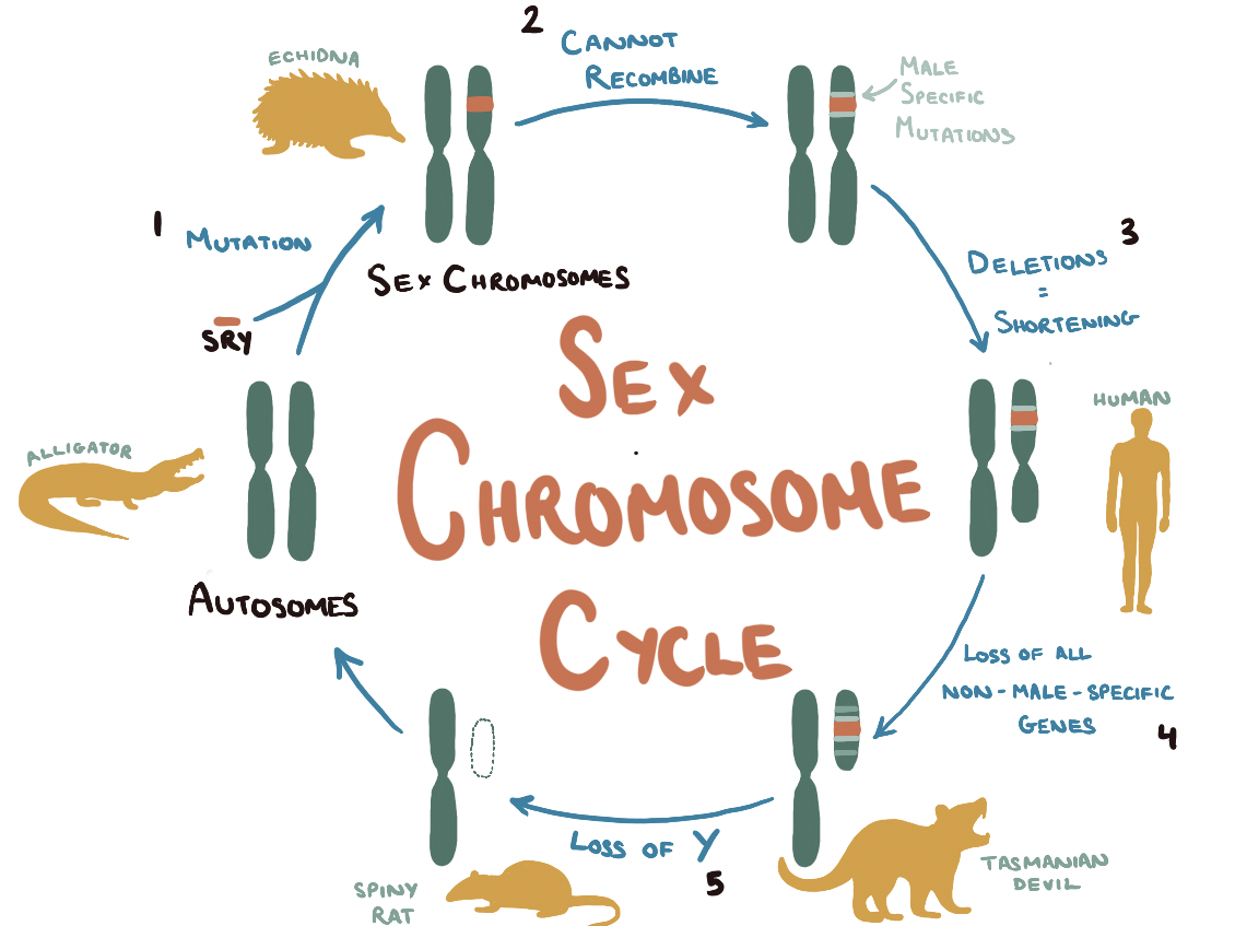
Prior to the evolution of mammals, sex determination did not follow a chromosomal blueprint. Rather it was decided by environmental factors. For example, alligators’ sex entirely depends on incubation and egg temperature at the time of hatching.  As a result, every one of their chromosomes belongs to a matching set as they lack distinctive sex chromosomes. At some point, very early in the evolution of mammals, an ancient form of the SOX3 gene, involved in brain development pathways, randomly mutated to form the SRY on a single autosome. The SRY gene codes for the testis-determining transcription factor (TDF) which binds to DNA to start a cascade of biochemical reactions leading to the differentiation of sexless primordial gonad cells into the sperm-producing cells of the testes. 

The switch from SOX3 to SRY was the first step in a long path of mutations, a growing divide between the SRY-containing autosome and its counterpart—culminating in two very different sex chromosomes. From comparing our genomes to those of early mammals, such as marsupials, geneticists have identified that the modern Y chromosome shares just four genes with its much larger autosomal ancestor. Due to the sex chromosomes inability to undergo recombination, the Y chromosome has gradually acquired a slew of deleterious mutations—filling the chromosome with ‘junk’ DNA and shortening it through deletion mutations. As the Y chromosome mutates, it becomes less able to recombine with its X counterpart and continues to lose genes resulting in a vicious cycle of shrinkage.

Dasyurid marsupials, such as the Tasmanian devil, have a Y that has been whittled down to just 10 megabases in length—a little genetic fragment containing just SRY and a few sperm-producing genes. This degradation is taken to the extreme in bandicoots. These marsupials use their miniscule Y chromosome (holding nothing but SRY, the last in a long line of seemingly disposable genes) for sex-differentiation and gonad formation, before eliminating it entirely from all other embryonic cells. Such a small chromosome, essentially a single gene, is at great risk of further loss. 

Only two species have ventured beyond this point in chromosomal evolution: the ryukyu spiny rat and the transcaucasian mole vole. Their Y chromosome has diminished to the point where it has been taken up by its X counterpart through the process of translocation. Now instead of XX females and XY males, the sex-determination system is XX females and XX’ males (where X’ is an identical X chromosome with an addition of the SRY gene). 

The X’ will not be able to entirely recombine with the X and gradually it will accumulate mutations—diverging in function and shortening to produce a new Y chromosome. In this way, the same Y chromosome evolution described earlier can renew itself cyclically in a sex chromosome cycle.

Cats Are Not Peas is a great read which explains many of the basic genetics concepts learnt in Year 1 of the MBiochem course such as X-inactivation, masking, variable gene expression, polygenes, mosaicism and chimerism. The book doesn’t include many recent developments in genetics (such as sequencing technologies) as it was written pre-Human Genome Project in 1996. However, it still gives an interesting perspective on genetics research and the history of the field up until that point. It also piqued my interest in X inactivation and Y chromosome shortening and served as a starting point for further research. 


References:

Gould, L. (1996). Cats are not Peas. New York, Ny Springer New York.

Avner, P. and Heard, E. (2001). X-chromosome inactivation: counting, choice and initiation. Nature Reviews Genetics, 2(1), pp.59–67.

Morey C, Avner P (2011) The Demoiselle of X-Inactivation: 50 Years Old and As Trendy and Mesmerising As Ever. 

PLOS Genetics 7(7): e1002212. https://doi.org/10.1371/journal.pgen.1002212

Kashimada, K. and P. Koopman. “Sry: the master switch in mammalian sex determination.” Development 137 (2010): 3921 – 3930.

HHMI BioInteractive. (n.d.). Evolution of the Y Chromosome. [online] Available at: https://www.biointeractive.org/classroom-resources/evolution-y-chromosome [Accessed 14 September 2021].

Matveevsky, Sergey et al. “Chromosomal Evolution in Mole Voles Ellobius (Cricetidae, Rodentia): Bizarre Sex Chromosomes, Variable Autosomes and Meiosis.” Genes vol. 8,11 306. 3 Nov. 2017, doi:10.3390/genes8110306
Benjamin L S Furman, David C H Metzger, Iulia Darolti, Alison E Wright, Benjamin A Sandkam, Pedro Almeida, Jacelyn J Shu, Judith E Mank, Sex Chromosome Evolution: So Many Exceptions to the Rules, Genome Biology and Evolution, Volume 12, Issue 6, June 2020, Pages 750–763, https://doi.org/10.1093/gbe/evaa081

Cahill Cycle: how our Liver helps us Exercise

Standard

The following is a tutorial essay written for the Cellular Biochemistry module – the writing style is pitched at university-level but can also be followed by A Level biology students.

If you’re curious about the intricacies of glucose synthesis in the liver or just want to see what an Oxford biochemical tutorial essay looks like – read on!


The glucose-alanine cycle, also known as the Cahill cycle, describes the metabolic pathway in which extrahepatic tissue (notably, the skeletal muscle) exports alanine to the liver, alanine is used as a substrate in gluconeogenesis, and nascent glucose is exported back from the liver to the other tissues.

ALANINE –> PYRUVATE

Under extreme catabolic conditions, such as fasting, intracellular protein hydrolysis rate exceeds the rate of its resynthesis – resulting in the liberation of free amino acids which can either be oxidised for energy (in the case of the branched chain amino acids, leucine, isoleucine, and valine) or transported out of skeletal muscle into the bloodstream for gluconeogenesis in the liver.

In the liver, alanine is first deaminated in a transamination reaction, catalysed by hepatic alanine aminotransferase, in which α-ketoglutarate acts as an amino group acceptor. Overall, this reaction is

Alanine + α-Ketoglutarate ⇄ Glutamate + Pyruvate.

Aminotransferases are cytosolic enzymes, particularly abundant in liver cells, which require the coenzyme pyridoxal phosphate (PLP) to be bound to the active site. Transamination of alanine produces pyruvate and glutamate. Glutamate can then be deaminated by glutamate dehydrogenase to reform α-ketoglutarate which can enter the Krebs cycle; this anaplerotic reaction also forms NH4+ which enters the urea cycle to be safely excreted.

PYRUVATE –> OXALOACETATE

The pyruvate can leave the Cahill cycle at this point and be oxidised for ATP production; in periods of fasting, however, it is energetically favourable to remain in the cycle. The conversion of pyruvate into oxaloacetate, catalysed by pyruvate carboxylase, is the next step in the Cahill cycle. The metabolic pathway encounters a physical barrier here since the necessary enzyme is localised in the mitochondrial matrix. The mitochondrial pyruvate carrier (MPC) complex, consisting of dimerised subunits MPC-1 and MPC-2, translocates pyruvate into the mitochondria.

Expression of MPC is under hormonal regulation; increase in circulating pancreatic glucagon during fasting upregulates expression of MPC on mitochondrial outer membranes. Glucagon binds to G protein-coupled receptors and activates adenylate cyclase which increases cAMP production. cAMP phosphorylates PKA which in turn activates the cAMP responsive element binding protein (CREB). CREB, a transcription factor, directly stimulates gluconeogenesis by binding the promoter of the MPC1 and MPC2 genes as well as the promoters of important enzymes involved later in the glucose-alanine cycle. This long-term regulation promotes gluconeogenesis during extended periods of fasting in which blood glucagon concentration is consistently high.

Once in the mitochondria, pyruvate is converted to oxaloacetate by pyruvate carboxylase. The enzyme requires a covalently-bonded prosthetic group, biotin, to act as a carrier of activated CO2. Pyruvate carboxylase consists of four identical subunits each made of four domains: a biotin carboxylase (BC) domain, a biotin carboxyl carrier protein (BCCP), a pyruvate carboxylase (PC) domain, and a PT domain which stabilises the tetramer

First, carbonic acid reacts with ATP to produce activated CO2. This CO2 is then transferred to the biotin attached to the BCCP subunit, releasing a orthophosphate molecule. Finally, the CO2 is transferred to a pyruvate molecule (catalysed in the active site of the PC subunit) to form oxaloacetate.  

This is the first irreversible, committed step of the gluconeogenesis pathway and, hence, is a key step for regulation. The energy charge of the cell plays a crucial role in activating pyruvate carboxylase – biotin cannot be carboxylated unless the allosteric regulator acetyl CoA is bound to the enzyme. If the energy charge in the liver cell is low, acetyl CoA concentration will be low and pyruvate will instead be decarboxylated to enter the TCA cycle, inhibiting gluconeogenesis. If acetyl CoA concentration is high, phosphate dehydrogenase (a glycolytic enzyme) is inhibited and pyruvate carboxylase activity is stimulated.

OXALOACETATE –> PHOSPHOENOLPYRUVATE (PEP)

The enzyme that catalyses the conversion of oxaloacetate to PEP is only present in the cytoplasm so the tricarboxylic acid must first be transported out of the mitochondria. The inner mitochondrial membrane is impermeable to oxaloacetate but not to malate; reduction to malate via malate dehydrogenase allows the molecule to be transported by the malate- α-ketoglutarate transporter. In the cytoplasm, malate is then oxidised by cytosolic malate dehydrogenase to reform oxaloacetate.

The malate shuttle serves another important purpose as it enables the transport of reducing equivalents across the outer mitochondrial membrane (which is impermeable to NADH). When malate is converted back to oxaloacetate, cytoplasmic NADH is formed which reduces substrates later in the gluconeogenic pathway.

Oxaloacetate is simultaneously decarboxylated and phosphorylated (using GTP) by phosphoenolpyruvate carboxykinase (PEPCK) to produce PEP. Metal ions, Mn2+ and Mg2+ act as cofactors and stabilise the several negative charges interacting in this reaction.

Cytosolic PEPCK gene expression is hormonally regulated by glucagon via cAMP (as described earlier) as well as by cortisol. This hormone diffuses directly through the phospholipid bilayer of hepatic cells and binds to the glucocorticoid receptor (GR) to form a cortisol-GR complex. The complex then translocates into the nucleus and binds the Glucocorticoid Response Element upstream of the PEPCK gene – activating it.  Insulin, on the other hand, downregulates PEPCK expression – inhibiting gluconeogenesis.

By carboxylating and subsequently decarboxylating pyruvate, the phosphorylation of the substrate (typically a very endergonic reaction) is made more favourable. Direct addition of a phosphoryl group has a high ΔG  ̊of +31 kJmol-1, making the reverse dephosphorylation reaction irreversible. Decarboxylation drives the gluconeogenic pathway forwards by bypassing this unfavourable step with a much lower ΔG  ̊of +0.8 kJmol-1.

PEP à FRUCTOSE-1,6-BISPHOSPHATE

The six following reactions are an exact reversal of the glycolytic pathway and are catalysed by glycolysis enzymes; this is possible as all these reactions are reversible and near equilibrium in hepatic cells – if the metabolite concentrations favour gluconeogenesis, PEP will be converted to fructose-1,6-bisphosphate via:

Phosphoenolpyruvate + H2O 2-phosphoglycerate

2-Phosphoglycerate 3-phosphoglycerate

3-Phosphoglycerate + ATP 1,3-bisphosphoglycerate + ADP

1,3-Bisphosphoglycerate + NADH + H+ glyceraldehyde 3-phosphate + NAD+ + Pi

Glyceraldehyde 3-phosphate dihydroxyacetone phosphate

Glyceraldehyde 3-phosphate + dihydroxyacetone phosphate fructose 1,6-bisphosphate

FRUCTOSE-1,6-BISPHOSPHATE –> FRUCTOSE-6-PHOSPHATE

The dephosphorylation of F16BP to F6P, mediated by fructose-1,6-phosphatase (FBPase) and Mg2+ ion cofactors, bypasses another irreversible step of the glycolytic pathway and is a key point of regulation in the gluconeogenic pathway. FBPase is a tetrameric enzyme with an AMP binding site as well as an active site; fructose-2,6-phosphate competitively inhibits FBPase by binding the active site and sterically inhibiting the substrate F16BP from binding whereas AMP inhibits FBPase by binding an allosteric site.

F26BP is an allosteric effector molecule that is not an intermediate of either pathway but regulates both glycolysis and gluconeogenesis. Intracellular concentration of F26BP is under hormonal control; glucagon decreases the concentration whereas insulin increases it. Glucagon stimulates cAMP which activates PKA which phosphorylates a serine residue in the active site of the bi-enzyme PFK-2/FBPase-2. Phosphorylation increases the phosphatase activity of the enzyme, converting F26BP to F6P, so the effector is unable to bind and inhibit FBPase – stimulating gluconeogenesis. Insulin, on the other hand, binds to specific membrane receptors and activates phosphoprotein phosphatase 2A (PP2A) which catalyses the dephosphorylation of PFK-2/FBPase-2. This allows the bi-enzyme to act as a kinase and increase F26BP concentration – inhibiting gluconeogenesis.

AMP acts as a non-competitive inhibitor; upon AMP ligation, hydrogen bonds holding the FBPase tetramer together are disrupted causing a 15  ̊ to 17  ̊ rotation of the upper dimer relative to the lower dimer which converts the enzyme from the R state to the inactive T state.

FBPase regulation plays a crucial role in survival for obligate hibernators. During hibernation, glycolysis must be prioritised so hepatic gluconeogenesis is inhibited. Respiration rate dramatically decreases, creating relatively anoxic conditions in liver tissue which inhibits FBPase by making it more sensitive to allosteric inhibitors (ex. AMP, ADP, Pi) and F26BP. FBPase affinity for its substrate also decreases through covalent modification; for instance, hibernating bats show a 75% decrease in K­M compared to euthermic bats.

GLUCOSE-6-PHOSPHATE –> GLUCOSE

Fructose-6-phosphate is readily converted to glucose-6-phosphate by the same glycolytic enzyme that catalyses the reverse reaction since the reaction is maintained close to equilibrium.

Standard gluconeogenesis terminate here in most tissues since glucose-6-phosphate is unable to readily diffuse out of cells and can be converted to glycogen for faster mobilisation during aerobic respiration. However, the interorgan glucose-alanine cycle is unique in its production of free glucose which is transported out of the liver cells and recirculated to the skeletal muscle. This dephosphorylation requires a third bypass of an irreversible step of the glycolytic pathway; glucose-6-phosphatase, a protein complex embedded in the ER, catalyses the reaction instead of hexokinase/glucokinase. Only tissues that maintain blood-glucose homeostasis, such as the liver and kidney, contain these G6Pase complexes.

SUMMARY

The overall reaction for the conversion of alanine to glucose is:

2 Alanine + 10 ATP + CO2 Glucose + urea + 10 ADP + 10 Pi

The alanine-glucose Cahill cycle repurposes carbon skeletons of the liberated amino acid and provides a means of safely exporting and processing amino groups (via the linked Urea cycle) whilst ensuring that the liver bears the energetic burden of gluconeogenesis instead of actively respiring skeletal muscle tissue.

The three main points of regulation in gluconeogenesis:

  • pyruvate –> oxaloacetate
  • oxaloacetate –> PEP
  • F16BP –> F6P

correspond with irreversible steps in the glycolytic pathway. The main forms of regulation are hormonal control via antagonistic glucagon/cortisol and insulin as well as allosteric regulation by acetyl CoA and AMP.


BIBLIOGRAPHY

Lou, M., Li, J., Cheng, Y., Xiao, N., Ma, G., Li, P., Liu, B., Liu, Q. and Qi, L. (2019). Glucagon up‐regulates hepatic mitochondrial pyruvate carrier 1 through cAMP‐responsive element‐binding protein; inhibition of hepatic gluconeogenesis by ginsenoside Rb1. British Journal of Pharmacology, 176(16), pp.2962–2976.

Biochemistry, 4th Edition by Donald Voet, Judith G. Voet

Lubert Stryer, Berg, J., Tymoczko, J. and Gatto, G. (2015). Biochemistry. New York Macmillan Learning Wh Freeman.

Kaur, R., Dahiya, L., & Kumar, M. (2017). Fructose-1,6-bisphosphatase inhibitors: A new valid approach for management of type 2 diabetes mellitus. European Journal of Medicinal Chemistry, 141, 473–505. doi:10.1016/j.ejmech.2017.09.029 

Nelson, D.L., Lehninger, A.L., Cox, M.M., Osgood, M. and Ocorr, K. (2009). Lehninger principles of biochemistry. New York: W.H. Freeman

Felig, P., Pozefsk, T., Marlis, E., & Cahill, G. F. (1970). Alanine: Key Role in Gluconeogenesis. Science, 167(3920), 1003–1004. doi:10.1126/science.167.3920.1003 

N, S. (2019). What is Gluconeogenesis? Definition, Steps, Substrates & Regulation. [online] Biology Reader. Available at: https://biologyreader.com/gluconeogenesis.html [Accessed 30 Jan. 2021].

A Sixth Sense – the science behind Migraines with Aura

Standard

So Much More Than ‘Just A Headache’

Unfamiliar with the extreme pain a migraine brings, it’s natural to try and relate it to a bad headache – mistaking the condition’s throbbing pain for a common distracting ache. Besides intense pain, migraines also come with a host of sensory issues such as severe sensitivity to sound and light (known as photophobia), neck stiffness, and visual phenomena. While headaches are an infamous symptom of illness, migraines are the illness.

The most striking difference between a migraine and a headache is that, almost as if they have a sixth sense, one in three migraine sufferers can predict when one is brewing before the pain sets in. Relax – we’re not omniscient. In the build up to a migraine attack, we experience auras: transient visual disturbances that are as disorientating as they are fascinating from a scientific perspective.

What starts out as a shimmering haze in the migraine sufferer’s periphery, gradually envelops their field of vision with flashing lights, shimmering arcs, and blind spots. Although auras serve as a warning for an impending migraine attack, they can be far more dangerous than the ensuing headache; during auras, brain cells can become damaged, even to the point of death, causing ischaemic strokes and creating lesions in the white matter of the brain. But what’s the connection between a migraine and these strange visual symptoms?

Origin of an Aura

At the onset of a migraine, the biochemistry of the brain changes drastically. At rest, healthy neurons contain fewer positively charged ions such as sodium ions (Na+) and potassium ions (K+) and more negatively charged proteins and nucleic acids than their surroundings. This separation of charge gives their membranes a low resting potential of -70 mV.

If we compare a neuron’s axon to a simple tube connecting one neuron to its neighbour, we can see that the inside of the axon is more negative than its surroundings outside the cell (the extracellular space).

Triggering the migraine aura is a sudden, spontaneous efflux of K+ ions out of a single neuron buried deep in the brain’s visual cortex – causing the cell’s membrane potential to plummet. The build-up of K+ ions in the extracellular space triggers the release of amino acid glutamate which excites neighbouring neurons and allows K+ ions to flood in – propagating the ion movement further. On the heels of this sudden flurry of activity, comes a wave of neuronal silence in which each cell stops processing and transferring information as it slowly returns to normal.

This wave of intense excitement followed by inactivity spreads across the visual cortex – corrupting visual information from the eyes and creating strange auras.

This phenomenon, known as cortical spreading depression (CSD), is responsible for the peculiar auras that slowly take over a migraineur’s visual field. When a neuron is affected by the spreading depression, it stops accurately processing visual information from the eyes – instead interpreting signals into disturbances like flashing lights or blurry blind spots.

The gradual spread of CSD across the visual cortex is mirrored by the gradual growth of an aura in the build up to a migraine attack.

Cortical spreading depression is not unique to the visual cortex; it can occur throughout the grey matter. The reason why this wave of electrical activity originates in the visual cortex is because this region of the brain is very densely crammed with neurons – increasing the risk of one neuron’s aberrant activity affecting the many cells around it.

CSD & Pain

The effects of CSD extend far beyond interfering with vision. Neuron excitation in the visual cortex activates adjacent trigeminal nerves which span your face from temple to jaw. These nerves run parallel to major blood vessels of the meninges (the cushioning tissue between your skull and brain) and, when activated, trigger the release of pain-generating substances into the bloodstream. As a result, the meninges become inflamed, causing blood vessels to widen and put further pressure on the tissue – intensifying the pain.

To make matters worse, these waves of neural activity up-regulate the expression of genes encoding pro-inflammatory molecules (such as COX-2, the target of many painkillers) exacerbating the sensation of pain. Additionally, CSD activates metalloproteinases, provoking the blood-brain barrier to leak; pro-inflammatory molecules, as well as the K+ and glutamate from earlier, are now free to flood into the major facial blood vessels. In combination, these molecules sensitise the nerves in the neck and jaw, causing the notorious aches and stiffness many migraine sufferers endure. This sensitisation is also why normally innocuous processes (like coughing and yawning) hurt and cause tension to build up during a migraine.

The spreading depression is first transmitted to the ophthalmic nerve branch and is then propagated to the mandibular nerves, bringing the pain along with it.

Potential Treatments

None of these excruciating effects are felt in ordinary headaches where CSD plays no role; to reiterate, the intense pain felt by migraine sufferers comes down to the unique biology of a migraine attack – it has nothing to do with low pain thresholds and its far more than just a bad headache.

Currently, the majority of migraine treatments either suppress pro-inflammatory molecule production or tackle the excessive blood vessel widening which puts pressure on the meninges. While they are useful in dealing with the headache aspect of a migraine attack, they have no effect on auras and fail to address the root cause of the migraine: the CSD.

Drugs that interfere with dopamine and serotonin pathways have been proven effective, quite possibly because they control neurotransmitter release and hence limit excitation of adjacent neurons in CSD – curbing the spread of depolarisation. Potential novel treatments also address the K+ ion channel dysfunction that triggers CSD, preventing both the aura and the pain that follows it.

When it comes to migraine treatment, the classic proverb rings true – prevention is better than cure. However, there are shockingly few effective migraine prophylactics.
Research into what triggers the K+ ion efflux of CSD in the first place and why sufferers are genetically predisposed to having migraines could spare the nearly 10 million of us in the UK from frequent bouts of agony.

.

Bibliography:

  • Pietrobon, D., Striessnig, J. Neurobiology of migraine. Nat Rev Neurosci 4, 386–398 (2003). https://doi.org/10.1038/nrn1102
  • Lauritzen, M. (1987). Cortical spreading depression as a putative migraine mechanism. Trends in Neurosciences, 10(1), 8–13. doi:10.1016/0166-2236(87)90115-9 
  • Charles, A. C., & Baca, S. M. (2013). Cortical spreading depression and migraine. Nature Reviews Neurology, 9(11), 637–644. doi:10.1038/nrneurol.2013.192 
  • Medscape.com. (2019). Migraine Headache: Practice Essentials, Background, Pathophysiology. [online] Available at: https://emedicine.medscape.com/article/1142556-overview#a3.
  • Spierings, E. L. H. (2001). MECHANISM OF MIGRAINE AND ACTION OF ANTIMIGRAINE MEDICATIONS*. Medical Clinics of North America, 85(4), 943–958. doi:10.1016/s0025-7125(05)70352-7 

I’ve written this post for Migraine Awareness Week – if you wish to support research into treatments for this debilitating neurological condition, consider donating to Brain Research UK.

The Most Important Race of Your Life

Standard

Before the first cells of our bodies are even formed, we all owe our lives to electricity. It plays a crucial role in the first and most important race of our lives, regardless of whether we grow up to be marathon runners. This is the race of fertilisation, in which the finish line is an egg cell and the winner is the sperm cell that made each one of us.

In the same way that professional athletes have light, aerodynamic bodies and strong legs to propel them, sperm cells are streamlined and specialised to win their race. But instead of powering their movement with muscular limbs, sperm cells have powerful tails, known as flagella, which beat from side to side, tunnelling their way through the viscous fluid of the female reproductive tract.

At first, the race is akin to a marathon in which covering the distance between the vaginal opening and the uterus, without running out of energy, is the main priority. Dyenins, molecular motors embedded in the base of the flagella, produce a rapid symmetrical bending movement which quickly move the sperm cells to the upper reproductive tract in under half an hour.

From Ions to Energy
Just like the motors which keep the blades of ceiling fans spinning, these molecular motors need a constant supply of energy to keep the flagella beating. This energy comes in the form of tiny molecules called ATP which are continuously pumped out of miniature factories known as mitochondria. Mitochondria are bean-shaped organelles which have two membranes – an outer membrane which covers the organelle like a skin and a highly-folded inner membrane keeps important chemicals inside the organelle. There are mitochondria in all the cells in our bodies, but sperm cells need so much ATP to swim all the way to the egg that they are absolutely brimming with them. To produce all that ATP, mitochondria themselves require electrical energy.

So how do they get this electrical energy? First, let’s quickly return to our analogy of the ceiling fan, which uses electrical energy directly. Flick the switch on and extremely tiny charged particles flow through the wires of the fan motor. Electricity is really just the flow of any charged particle; when electricity flows, energy transfers along with it. The device you are reading this on is powered by the flow of negatively charged electrons. Our bodies, on the other hand, are mainly driven by slightly larger, positively charged particles: ions.

Our mitochondria are extra special because they can make use of both ions and electrons. First, these miniature factories shuttle around electrons to create flows of charge: bioelectricity. The resulting energy transfer is used to push positively charged hydrogen ions (H+) out of the mitochondrial inner membrane into the empty space between membranes. This causes a build-up of H+ ions on one side of the inner membrane and a loss of H+ ions on the other side. This results in one side of the inner membrane being more positively charged than the other – creating an electrical potential gradient (aka a voltage).

As I write this article, my abandoned cup of piping-hot tea is quickly turning cold; in the same way that heat tends to flow from a hot object to the cold surrounding air, charged particles tend to move from a positively charged area to a negatively charged one, down to the electric potential gradient. H+ ions in the mitochondria’s intermembrane space try to squeeze back through the inner membrane to the more negatively charged side.

But biological membranes repel H+ ions because they are charged so the ions cannot pass through freely. Mitochondria take advantage of that fact by having lots of specialised ion channels embedded in their inner membrane; these channels allow H+ ions to easily diffuse through to the more negatively charged side. But there is a catch. As H+ ions flow through the specialised channels, their electrical potential energy is transferred to the channel and used to manufacture more ATP!

We have now seen two ways sperm use bioelectricity, in their mitochondria, to help spin their flagella. Bioelectricity is essential for the repetitive side-to-side beating needed to get the sperm to the uterus. But the sperm cell’s journey doesn’t end there…

A Sticky Situation
When a sperm cell draws nearer to the egg cell, its marathon transforms into an obstacle course. Before it can even begin to navigate the sharp turns and swooping bends of the fallopian tubes, the sperm gets trapped by spindly finger-like projections at the tube entrance.

To free itself from the clutches of the upper reproductive tract, a change of movement is required. Sperm cells don’t have another type of molecular motor which takes over the flagella, but they do have a trick up their metaphorical sleeves, and this too involves bioelectricity.

As the sperm cell travels through the uterus, the environment changes. Earlier, the cell journeyed through a mixture of seminal and cervical fluid. Now, as it reaches the fallopian tube, it must swim through the sticky fluid released by the ovaries. Known as follicular fluid, this stuff is far richer in positively charged sodium (Na+) ions than the sperm cell’s cytoplasm. As a result, there’ is an imbalance of charge between the inside and outside of the sperm cell: a voltage .

This voltage triggers the opening of specialised exchanger proteins in the sperm cell membrane which can trade sodium ions outside the cell for hydrogen ions within the cell. As the sperm cell pumps out H+ ions through this exchanger, the concentration of H+ ions rapidly plummets. The pH of the cell’s cytoplasm is inversely proportional to H+ concentration; as the ion concentrations decreases, pH increases – making the cytoplasm more alkaline. This is crucial for initiating the next phase of sperm movement.

Modifying Movement

While the sperm cell is still held hostage by the spindly projections of the fallopian tube, it gets bathed in even more follicular fluid, which is rich in the hormone progesterone. Sperm cells are highly receptive to even a trillionth of an increase in progesterone concentration, so this flood of hormone induces great changes in its behaviour.

In combination with the alkaline environment, progesterone triggers the opening of CatSper channels (Cationic channels of Sperm) which allow positively charged calcium ions to flood into the sperm, yet again modifying the voltage across the sperm cell membrane.

More importantly, these calcium ions influence the dyenins in sperm flagella, so they produce an asymmetrical, whip-like movement. Adding calcium ions is akin to adding gears between the motor and the wheels of a car when you need more power to drive up a steep hill. The Ca2+ ions slow down the molecular motors and increases the overall bending of the flagella so that each beat of the sperm’s tail is more forceful. With this extra power, sperm cells can kick away from the clutches of the fallopian tube entrance. The asymmetric movement, combined with the sperm cell’s attraction to progesterone, allow it to steer a course towards the egg cell.

Bioelectricity, in the form of changing voltages and positive charge flow, is vital for sperm cells to break free from the microvilli projections at the entrance and make that final push through the fallopian tubes to the finish line of fertilisation. Staggeringly few sperm cells make it. Only one sperm cell out of every million that enter the female reproductive tract even makes it to the fallopian tube and less than a hundred ever come close to the egg. To put this in context, a healthy man can ejaculate about a teaspoon of semen, teeming with 500 million sperm cells – each sperm has a shockingly slim 0.00002% chance of even meeting the egg.

Without the powerful whipping motion that this bioelectricity enables, sperm cells can’t fertilise an egg cell and so a male cannot transfer genetic material to produce offspring. Ultimately, bioelectricity issues can result in infertility, a condition that affects 7 in 10 British couples.

Polyspermery: It’s A Draw
The converse issue, wherein lots of sperm cells are very efficient swimmers and reach the egg cell at the exact same time, also hinders reproduction. In a phenomenon known as polyspermy, two or more sperm cells fertilise the egg cell at the same. In terms of our race metaphor, polyspermy happens when two competing sperm cells ‘draw’ and there is no winner.

An egg cell fertilised by two or more sperm cells cannot divide the contributed genes evenly to split into new cells and produce an embryo, so the fertilised cell dies. As we’ll see shortly, polyspermy isn’t a major threat for humans since multiple sperm cells rarely reach the egg at the exact same instant. However, the risk of polyspermy is a serious issue for many marine animals that rely on external fertilisation to form offspring.

Sea Urchin Sex
Consider sea urchins, probably the spikiest creatures beneath the waves. Male and female sea urchins reproduce by releasing clouds of sperm and egg cells into the water and hoping they fuse to form offspring larvae. Compared to humans, many more sea urchin sperm cells approach a single egg simultaneously. To prevent polyspermy, a fertilised egg cell needs to act fast and block access to any more competitor sperm. This calls for bioelectricity.

An urchin egg cell waiting to be fertilised contains fewer positively charged ions than the surrounding seawater. This disparity in positive charge between its internal and external environment create a voltage known as a membrane potential; in the case of an unfertilised sea urchin egg cell, this membrane potential is -70 mV.

The very instant that the first sperm cell, the winner of the fertilisation race, binds to the egg cell, it triggers calcium and sodium ion channels to open and allow the positively charged ions to flood in. This results in a reversal of membrane potential as the inside of the egg cell becomes more positive than its surroundings, depolarising the membrane to +25 mV.

This rapid switch in membrane charge inhibits additional sperm cells from binding, however the exact mechanism remains a mystery. One prevailing theory is that a positive membrane potential inactivates the specialised slots in the egg cell membrane that allow sperm cells to dock in – preventing additional sperm from fusing with an already fertilised egg.

Alternatively, the positively charged proteins on the tip of the sperm cell, which fuse it to an egg, might be repelled by the newly positive membrane of the egg cell, ultimately inhibiting fusion and polyspermy. Regardless of the true mechanism, it is evident that bioelectricity plays an important role in guaranteeing successful fertilisation, and not only in sea urchins.

Slow Blocks
Polyspermy is not as immediate a threat to fertilisation for humans due to the relatively low sperm-to-egg ratio and the complexities of navigating the female reproductive tract. Instead of a fast, electrical block, our fertilised egg cells block out additional sperm through chemical methods: a slow block . To understand how human egg cells do this, we need to first look at its structure and its vulnerability to polyspermy.

Like all body cells, egg cells have a standard membrane that keeps all its cellular machinery and DNA inside, safe and secure. On top of this, egg cells are coated in a transparent jelly layer called the zona pellucida, which allows sperm cells to tether to the egg during fertilisation. The slow block to polyspermy involves changes in both the inner membrane and the zona pellucida.

Within minutes of fertilisation, the ‘winning’ sperm triggers calcium ions from within the egg cell to be released from their storage spaces and rush through the cell membrane. But unlike in the sea urchin eggs, this ion movement is not a one-time event for human egg cells. In the next minute, another wave of positively charged ions are sent out, and this whole process is repeated over and over and over. The end result is the formation of a fluctuating membrane potential which steadily flicks from positive to negative like a metronome.

These calcium ion oscillations themselves do not deter additional sperm. Instead, they orchestrate a cascade of reactions which trigger little pockets of enzymes to be pushed out of the egg cell. The enzymes inside can then dissolve the bonds between the membrane and the zona pellucida, pushing away the jelly layer and all the other attached competitor sperm cells along with it.

An IVF Issue
For natural human conception, this slow block is sufficient. However, within the last fifty years, this process has been unable to keep up with a novel form of reproduction we have pioneered: in vitro fertilisation (IVF). The conditions of IVF are very different to that of natural conception – the reproductive tract with its pH gradient, microvilli obstacles and sharp bend is swapped out for a simple laboratory dish. Many more sperm cell are incubated with a single egg to increase the chance of successful fertilisation. The consequence is that, when the playing field for sperm is levelled to this extent, the classic slow block might not act fast enough to block out additional sperm cells. Up to 10% of fertilised eggs in IVF become non-viable due to polyspermy.

Recent studies suggest that egg cell quality could influence polyspermy block speed. By studying our rodent friends, biochemists have found that older mice eggs are more likely to have abnormal calcium signalling. The oscillations are irregular, like a broken metronome swaying wildly out of time. As a result, not enough enzyme pockets are recruited to the membrane. This means that the zona pellucida does not completely detach from the egg cell – allowing some sperm cells to stay stuck in place and squeeze inside.

Moreover, the in vitro setting and egg freezing involved in IVF decrease both the frequency and duration of calcium ion oscillations. Whilst screening eggs for their quality is still out of reach, bioelectricity research could improve egg preservation techniques – bringing the finish line closer in the most important race of our lives.

Through the Looking Glass

Standard

Through The Looking Glass

Lewis Carroll’s sequel to Alice’s Adventures in Wonderland was a firm favourite of mine in childhood. After all these years, memories of the mirror world Alice falls into come flooding back to me as I study organic chemistry.

In Through the Looking Glass, initially the mirror-world Alice explores is almost exactly the same as the world we are all familiar with except it’s a mirror image. For instance, Alice opens a book of poems and expects to see the words

j2

but is astounded to instead find written

j1

The words on her page are mirror images of each other but, within these words, some of the letters appear identical to what Alice expected to find (A, W, & O). An ‘A’ from the mirror-world is a superimposable mirror-image of an ‘A’ from the regular world. For some letters, however, this is certainly not the case; J, B, E, R, C, and K are not identical to their mirror-world counterparts. These letters are non-superimposable mirror images.

Now let’s imagine an even stranger world where the word ‘JABBERWOCKY’ is arranged like this:

j3

In this three-dimensional arrangement, these little groups of letters are bonded together and frozen in this specific spatial arrangement with ‘C’ protruding out of the page and ‘KY’ extending back into the page.

In a mirror-world, we would expect to find the mirror image of this strange 3-D word:

j4

These two 3-D words are mirror images of each other but they are non-superimposable. Even if Alice were to reach into the page and twist the ‘mirror-world’ word around by 180 degrees, it would not be identical to our original 3-D word since the ‘C’ would be extending back into the page instead of protruding out of it.

j5

mirror-world rotation

j3

original

instead of

Hence, the original 3-D word and the mirror-world word are non-superimposable mirror images. This Lewis Carroll analogy has been stretched to its limits so let’s return the realm of organic chemistry…

Many organic compounds also have non-superimposable mirror-image molecules. Such pairs of molecule are known as enantiomers. These doppelgangers are optical isomers since they differ in terms of spatial arrangement and not atom connectivity. Optical isomerism can arise from the fascinating concept of chirality. Chiral molecules possess at least one carbon atom bonded to four different groups (a chiral centre) and are not identical to their mirror image molecule.

Any molecule which exhibits reflective symmetry is, by definition, achiral; this means that all the amazing asymmetrical molecules that make up biological systems are chiral – opening up new avenues of complexity and complications.

When chiral molecules are synthesised in the laboratory, the specific enantiomer formed is determined by random chance and so chemists usually end up with a 50/50 split of the two enantiomers; this mixture, known as a racemate, is chemically pure but has different optical properties and different interactions in biological systems – ushering in a plethora of unforeseen obstacles and opportunities.

Drugs with Doppelgangers

All the components of living systems, right down to enzyme and receptor molecules, are chiral and so can only interact with one of the enantiomers of any pair of molecular twins. Therefore, the ‘active’ enantiomer of a drug has a three-dimensional structure that perfectly aligns with the binding site whereas the inactive enantiomer cannot bind in the same way no matter how much you rotate it.

Chiral carbon & chiral drugs | Stereochemistry (article) | Khan ...

A great example of this is epinephrine, more commonly known as adrenaline; the receptor site on the post-synaptic membrane of a neuron contains three binding sites complementary to the three functional groups of the more active enantiomer. Firstly, there is an anionic site which can form electrostatic forces of attraction with the positively charged ammonium ion group. At the opposite end of the molecule, the phenolic ring is attracted to the flat site of the receptor with π-π interactions.Receptor

The final receptor site can form hydogen bonds with the β-hydroxyl group attached to the chiral carbon in the active enantiomer of epinephrine. The inactive enantiomer, on the other hand, has a different orientation of the β-hydroxyl group so cannot form this crucial intermolecular bond. Hence, this enantiomer only has two points of contact with the receptor molecule – resulting in a hundred-fold decrease in activity which makes it ‘inactive’.

Each molecule in a synthetic epinephrine racemate has a 50/50 chance of being the active enantiomer and, thus, an effective neurotransmitter which means that higher doses are needed to achieve a desirable outcome such as an increase in post-synaptic action potentials. Needless to say, organisms would waste far too much energy synthesising biological molecules if only half of them could be metabolically active. Otherwise, for both enantiomers to be catalysed, our DNA would need to code for twice as many enzymes and receptor proteins which is similarly inefficient. In order to overcome this issue of ensuring complementary chirality, life plays by an entirely different set of rules.

The Principle of Homochirality

All life forms on earth exclusively synthesise and use L-amino acids and D-sugars so all naturally-occurring molecules are enantiomerically pure. This principle of homochirality can be explained from a biological standpoint by turning back the hands of time to the point where life first starting flourishing on earth.

When early life was first evolving, the enzymes synthesised by a microorganism would have been complementary to either the L-enantiomer or D-enantiomer, however the microorganism’s environment would have consisted mainly of one enatiomer or the other. The only microorganisms that survived and multiplied in our early environment would be ones that produces enzymes complementary to the most prevalent enantiomer of any molecule. Since early microorganisms were in environments abundant in D-sugars and L-amino acids, selective pressures would give rise to populations of microorganisms with enzymes and receptors only complementary to these enantiomers. Hence, every subsequent organism evolved from these early life forms would also be force to use the same amino acid and sugar enantiomers as its predecessors: resulting in universal enantiomer preferences.

cap2

There is a straightforward reason as to why D-sugars are more abundant than their L-enantiomer counterparts; sugars can spontaneously undergo cyclisation reactions to form the ring structures we are more familiar with. D-sugars form more stable rings than L-sugars and so are more prevalent in our environment.

fjgdg

circularly polarised light

The reason for the prevalence of L-amino acid is less intuitive and requires us to venture even further back in time to before life even began. Chirality doesn’t just exist at a molecular level; our galaxy itself rotates with a chiral spin which causes cosmic dust to circularly polarise the light travelling through space. From studying amino acid residue in meteors and comets, we can ascertain that circularly polarised light degrades D-amino acids to a greater extent than L-amino acids, resulting in the greater stability of L-amino acids.

This discrepancy in amino acid degradation, however, is not sufficient to explain the exclusive presence of L-amino acids in biological systems. Hence, we must consider the biochemistry behind the formation of chiral compounds.

In a primordial mixture of amino acids on earth, L-enantiomers act as auto-catalysts and catalyse their own formation, increasing the rate of L-enantiomer formation with increasing concentrations. Additionally, L-enantiomers can bind to D-enantiomers in a process known as mutual antagonism which inhibits auto-catalysis and further formation of the D-enantiomer. Auto-catalysis of L-amino acids, in combination with mutual antagonism, serve to exacerbate the discrepancy in amino acid ‘survival’, contributing to the favouring of L-amino acids over D-amino acids on our planet.

Isomer Inversion

ibup

Although almost all receptor and enzyme active sites in the body are only complementary to L-amino acids and D-sugars, certain enzymes, known as racemases, can convert a molecule’s inactive twin into the appropriate enantiomer in the process of stereochemical inversion. AMACR, for instance, converts a range of molecules from the R-enantiomer to the S-enantiomer.

One such molecule is the nation’s favourite painkiller: ibuprofen. Ibuprofen is administered as a racemate but undergoes a series of changes in the body (catalysed by AMACR) which convert all the R-ibuprofen to S-Ibuprofen which can inhibit cyclooxygenases and reduce the production of prostaglandins – hence creating an anti-inflammatory effect.

Increase in AMACR activity above certain threshold is hallmark of prostate cancer as AMACR is used to invert the chirality of molecules involved in accelerated cell proliferation and tumour growth. A prospective treatment of prostate cancer involves administering ibuprofen to prostate cells so that excess AMACR will catalyse the formation of S-Ibuprofen instead of molecules associated with tumour growth, resulting in a chemopreventive effect.

Mirror Milk

‘How would you like to live in Looking-glass House, Kitty?

I wonder if they’d give you milk in there?

Perhaps Looking-glass milk isn’t good to drink…’

In Through the Looking Glass, Alice raises the concern of her cat drinking looking-glass milk. From what we now understand of chirality, it is safe to say that neither the cat, nor Alice herself, would fare well in a mirror-world where their digestive enzymes cannot break down milk which is rich in D-amino acids an L-sugars (although the fats in milk, which are all achiral and therefore unchanged, could provide them with enough energy to make a quick escape).

But perhaps Kitty’s enantiomeric twin produces enzyme and receptor molecules proficient in synthesising and utilising these strange enantiomers.

Perhaps mirror-cats are partial to mirror-milk

Capture


Bibliography:

Part IV: The Future of Medical Scanning

Standard

The future of medical scanning is prognostic; hyperpolarised MRI scans are very promising since they can identify trace levels of biomarkers indicating future disease. Carbon-13-enriched glucose can act as a tracer to detect abnormal metabolism characteristic of burgeoning Type-2 diabetes. Para-hydrogenated bicarbonate ions can be injected to examine microenvironment pH levels suggestive  chronic inflammation.

Preemptive molecular scanning has the potential to form a prognosis before symptoms even appear – saving patients from years of suffering.

Another new avenue of development in the medical scanning sector that is gaining traction is the utilisation of artificial intelligence (AI) in analysing scans. Currently, a large wealth of information can be yielded from a single patient visit; however, trawling through vast amounts of data makes the diagnostic process time-consuming and labour intensive – increasing the risk of misjudgement and oversight.

Additionally, at the moment, a large proportion of valuable scan data is eventually discarded. This presents a significant waste of Big Data which could be repurposed for training the neural networks of AI systems to recognise subtle patterns in a scan, hence supporting faster and more reliable diagnoses.

Within the last decades, imaging techniques are being rapidly perfected and fine tuned even for extremely niche requirements. The rapid emergence of new technology and the tremendous advancement in medical scanning is epitomised by the very recent development of the highly-sensitive wearable MRI scanner which is adept at imaging the intricate interplay of bones, tendons and ligaments in motion; the image produced by this cutting-edge technology is reminiscent of the very first radiograph featuring the hand of Röntgen’s wife.

20180522-glove-combined.jpg
The juxtaposition of the two scans (taken just over a century apart) highlights the major strides made in the field of medical scanning.

In these scans, I do not see my death, as Röntgen’s wife did, but rather, the limitless potential for life-saving scanning technology.

Final Essay (1)


The figures quoted in this essay have been meticulously researched and documented. Below is the bibliography for this section of my essay:

“Super-Cool Imaging Technique Identifies Aggressive Tumors.” National Institute of
Biomedical Imaging and Bioengineering, National Institutes of Health, https://www.nibib.nih.gov/news-events/newsroom/super-cool-imaging-technique identifies-agressive-tumors [Last Accessed: 03 Mar 2019]

Zhang, Bei, Daniel K. Sodickson, and Martijn A. Cloos. “A high-impedance detector-array glove for magnetic resonance imaging of the hand.” Nature Biomedical Engineering 2 (2018): 570-577. Nature. Print. [Last Accessed: 25 Feb 2019]

Image Credit:

Zhang, Bei, Daniel K. Sodickson, and Martijn A. Cloos. MR image from wearable glove
coil design. Nature Biomedical Engineering, Nature, May 2018. Web.
https://www.researchgate.net/figure/Wearable-glove-coil-design-and-images-a-Photos-of-the-glove-coil-with-the-hand-stretched_fig4_324952145

Röntgen, Wilhelm. “Hand mit Ringen (Hand with Rings).” Wikipedia, Wikimedia
Foundation Inc., 22 December 1895

Part III: Nuclear Magnetic Resonance

Standard

As opposed to x-ray and ultrasound scanning devices, MRI uses the body’s innate magnetic properties to produce scans. Magnetic resonance imaging takes advantage of the high abundance of hydrogen nuclei (protons) in soft tissues of the body (which contain water and fat) and the fact that, often, diseases are manifested as an increase in water content as a natural consequence of inflammation.

Every hydrogen proton in the body has a spin and, hence, a small magnetic moment (1). An MRI machine applies a very strong magnetic field across the body to align all the hydrogen protons with the field (2). A radiofrequency pulse is then introduced which forces the hydrogen protons to realign against the magnetic field (3). Once the pulse is removed, the hydrogen protons return to their original alignment and release electromagnetic energy as MR signals which can be detected (4).

Final Essay

Tissue can be analysed and differentiated based on how much energy its constituent hydrogen protons release and how long it takes for them to return to their original  alignment (the relaxation time). Since the development of the first MRI scanner, several variants of the underlying principle have emerged to address niche situations.

BOLD fMRI
Blood-oxygen-level dependent functional MRI (BOLD fMRI) works on the principle that an increase in neuronal activity is always accompanied with an increase in oxygenated blood flow in local capillaries as the demand for oxygen increases.

The haemoglobin which delivers oxygen to the activated neurons is paramagnetic when deoxygenated (T-state) but diamagnetic when oxygenated (R-state); this unique magnetic property (caused by its different quaternary structures) results in slightly different electromagnetic emissions from the hydrogen protons within the haemoglobin depending on the level of oxygenation.

Screenshot 2019-09-19 at 1.40.20 PM

The MR signal can be analysed to infer a plethora of information – allowing clinicians to monitor the level of brain activity in a patient suffering from neurodegeneration and enabling researchers to discover the regions of the brain responsible for both instinctive reflexes and complex contemplation.

With its extreme versatility and minimal health risks, MRI arguably forms the heart of modern radiography – however, for certain patients with underlying ailments, the very scanning modality that is renowned for its safety can be deadly.

Safer Contrast Agents
Contrast agents are frequently used in MRI scans to alter the relaxation time of the hydrogen protons through their interaction with strongly paramagnetic gadolinium ions in the dye – this changes the MR signal intensities and increases the contrast of the scan. The gadolinium ions then accumulate in the kidneys for elimination. However, in cases of poor renal function, this can prove to be toxic. Patients with advanced chronic kidney disease, if injected with a typical gadolinium-based contrast agent, are at a significant risk of developing painful, debilitating disorders including contrast-induced nephropathy.

Just last year, a team at Massachusetts General Hospital devised a solution by proposing an alternative, safer manganese-based contrast agent (Mn-PyC3A) which not only produces identical contrast enhancement but is also made of an essential element which poses no risk to human health.

Hyperpolarisation
Within the past decades, although MRI has become indispensable in medical diagnostics, its sensitivity leaves much to be desired. In a typical MRI scan, due to the way that the molecular energies are naturally distributed, the MR signal from only 1 in 200,000 molecules is actually detected. Consequently, vital molecules (including potential cancer biomarkers) remain undetected due to their inherently low concentrations (<1mM) – severely restricting the scope of MRI.

Hyperpolarisation can overcome this sensitivity challenge by skewing the distribution of molecular energies and enabling detection of significantly more molecules. In hyperpolarisation, parahydrogen (polarised H2) or carbon-13 is chemically incorporated into an array of molecules (including glucose, urea, and pyruvate) and injected into the patient as tracers. This can increase the detected MR signal by up to 100,000 fold.

Hyperpolarisation drastically improves the imaging of metabolic activity – which is essential for tumour detection and monitoring. Cancer cells perform rapid glycolysis to obtain energy to fuel growth and division – converting pyruvate into the byproduct lactate in the process. A significant biomarker of aggressive, rapidly-growing tumours is a high lactate concentration. To assess the metabolic activity of a tumour, the patient is injected with carbon-13 enriched pyruvate and the concentration of hyperpolarised lactate produced is measured.

Unlike all other scanning methods, this technique yields information about not only the anatomy of a tumour but also its biochemistry – proving to be invaluable in assessing the efficacy of cancer treatment. Remarkably, within 24 hours of starting a new treatment, hyperpolarised MRI scans can reveal if a tumour is responding.

F6.large

The figure above shows a prostate tumour before and only 48 hours after starting treatment. The MRI scans in the first row show a slight decrease in tumour size whilst the graphs in the second row (which refer to the detected concentrations of lactate and pyruvate) show that the lactate concentration has plummeted – highlighting the success of the treatment.


This biochemical change is further illustrated in the lactate concentration maps (last row) generated from the hyperpolarised MRI scans.

With MRI hyperpolarisation, clinicians can swiftly identify the ideal treatment for each patient based on the unique metabolism of the tumour – representing a major milestone in advances toward personalised medicine.


The figures quoted in this essay have been meticulously researched and documented. Below is the bibliography for this section of my essay:

Berger, Abi. “How does it work?: Magnetic resonance imaging.” BMJ 324.7328 (2002): 35. PubMedCentral (PMC). Web. [Last Accessed: 21 Feb 2019]

NIBIB gov. “How Does an MRI Scan Work?” Youtube, 16 Apr 2013, https://www.youtube.com/watch?v=1CGzk-nV06g&feature=youtu.be [Last Accessed: 21 Feb 2019]

Devlin, Hannah. “Introduction to FMRI.” Nuffield Department of Clinical Neurosciences, University of Oxford, [Last Accessed: 22 Feb] https://www.ndcn.ox.ac.uk/divisions/fmrib/what-is-fmri/introduction-to-fmri 

“MRI contrast agent.” Wikipedia, Wikimedia Foundation Inc., [Last Accessed: 22 Feb 2019] https://en.wikipedia.org/wiki/MRI_contrast_agent

“Contrast Dye and the Kidneys.” A To Z Health Guide, National Kidney Foundation, https://www.kidney.org/atoz/content/Contrast-Dye-and-Kidneys [Last Accessed: 22 Feb 2019]

Gale, Eric, and Peter Caravan. “Gadolinium-Free Contrast Agents for Magnetic Resonance Imaging of the Central Nervous System.” ACS Chem. Neurosci. 9.3 (2018): 395-397. ACS Publications. Web. [Last Accessed: 22 Feb 2019]

Freeman, Tami. “Medical innovations: A safer MRI contrast agent.” physicsworld: Focus On Biomedical Physics. June 2018: 6. Print. [Last Accessed: 22 Feb 2019]

“What is hyperpolarisation?” Centre for Hyperpolarisation in Magnetic Resonance (CHyM), University of York, https://www.york.ac.uk/chym/hyperpolarisation/ [Last Accessed: 24 Feb 2019]

Miloushev, Vesselin, Kayvan Keshari, and Andrei Holodny. “Hyperpolarization MRI.” Top Magnetic Resonance Imaging 25.1 (2016): 31-37. PMC. Web. [Last Accessed: 03 Mar 2019]

Iali, Wissam, Peter J. Rayner, and Simon B. Duckett. “Using parahydrogen to hyperpolarize amines, amides, carboxylic acids, alcohols, phosphates, and carbonates.” Science Advances 4.1 (2018). American Association for the Advancement of Science. Web. [Last Accessed: 24 Feb 2019]

Ward, Christopher, et al. “Noninvasive Detection of Target Modulation following
Phosphatidylinositol 3-Kinase Inhibition Using Hyperpolarized 13C Magnetic Resonance Spectroscopy.” Cancer Research 70.4 (2010): 1296-1305. American Association for Cancer Research. Web. [Last Accessed: 04 Mar 2019]

“Super-Cool Imaging Technique Identifies Aggressive Tumors.” National Institute of Biomedical Imaging and Bioengineering, National Institutes of Health, https://www.nibib.nih.gov/news-events/newsroom/super-cool-imaging-technique identifies-agressive-tumors [Last Accessed: 03 Mar 2019]

“Cambridge extends world leading role for medical imaging with powerful new brain and body scanners.” Research – News, University of Cambridge, https://www.cam.ac.uk/research/news/cambridge-extends-world-leading-role-for medical-imaging-with-powerful-new-brain-and-body-scanners [Last Accessed: 03 Mar 2019]

Peirce, Andrea. “Hyperpolarized MRI: A New Tool to Assess Treatment Response within Days.” Memorial Sloan Kettering Cancer Centre, Sloan Kettering Institute,
https://www.mskcc.org/blog/hyperpolarized-mri-new-tool-assess-treatment-response within-days [Last Accessed: 03 Mar 2019]

Image Credits:

T-state: Liddington, R. “PDB ID 1HGA.” 1992. Originally published in the Journal of
Molecular Biology 228:551. Reproduced in Lehninger PRINCIPLES of BIOCHEMISTRY
(Seventh Edition). Print.

R-state: Silva, M. “PDB ID 1BBB.” 1992. Originally published in the Journal of
Biological Chemistry 267:17,248. Reproduced in Lehninger PRINCIPLES of BIOCHEMISTRY (Seventh Edition). Print.

Ward, Christopher, et al. “Figure 6. Effect of everolimus treatment on GS-2 tumour
xenografts” Cancer Research, American Association for Cancer Research, Web,
http://cancerres.aacrjournals.org/content/70/4/1296


Return to the Stamataki Lab

Standard

I’ve kicked off this summer with a trip to Birmingham to meet with Dr Zania Stamataki. During my prior work experience here, I improved my wet lab skills by learning essential techniques such as splitting cell lines, performing serial dilutions, and imaging plates with confocal microscopy.  This year, I chose to hone my computational biology skills to best prepare me for a future in research; Dr Scott Davies introduced me to bioimage informatics through training me to work with the ZEISS Efficient Navigation (ZEN) microscopy software.

The ZEN software can be used to process the plethora of data that a single imaged experiment can yield. I worked with data from an experiment in which Huh-7 hepatocytes (from an immortalised liver carcinoma cell line) are co-cultured with live T-cells and imaged using confocal microscopy.


I personally found this sophisticated mode of imaging fascinating so I did a little more research into the bioengineering responsible for the high resolution and vibrancy of the  cellular interactions captured by the confocal microscope. Unlike the humble optical microscopes of my school, which only need light and a pair of eyes to visualise cells, confocal microscopes require far more exotic elements: lasers and fluorescent dyes.


1gfl

GFP

One of the most widely used fluorescent biomarkers is green fluorescent protein (GFP) which is used in this experiment to highlight the cell membranes. GFP is a very important tool in research as it enables us to look beyond the cell surface and visualise organelles and protein movements. GFP has a unique sequence of the amino acids serine, tyrosine, and glycine which can be triggered to undergo chemical transformations in the presence of UV light. This special amino acid sequence is buried deep within the molecule’s barrel structure of intertwined protein sheets.


When activated, a cyclisation reaction occurs in which glycine forms a chemical bond with serine. This new closed amino acid ring spontaneously dehydrates and oxygen surrounding the GFP molecule forms a new double bond with the tyrosine – creating the fluorescent chromophore. In this way, GFP automatically assembles its own chromophore which can be used by researchers as tracking dyes. For instacnce, through genetic engineering, the GFP protein can be incorporated into the genome of specific cells or be used to label proteins of interest.  It is especially perfect for studying live cells since the alternative small fluorescent molecules (ex. fluorescein isothiocyanate) are highly phototoxic and inflict more damage upon live cells during imaging.

In the microscope images I worked with, GFP had been used to label the hepatocyte cell membranes. The T-cells were stained a vibrant blue whilst a cytoplasmic dye had rendered the hepatocyte cytoplasm red. The smaller dark regions enclosed within  cytoplasm are the hepatocyte nuclei.


Over the course of the week, I became proficient in producing orthogonal videos, which showcase the full depth of the hepatocytes, from multiple micrographs in a time lapse. Whilst processing these videos, I witnessed countless interesting phenomena; with Dr Stamataki’s permission, I’d like to share a few of my favourites with you.

Binucleate Hepatocyte Division

When I first starting working with the experiment data, Zania pointed this event out to me and I was both riveted and shaken to my core; this cell defied everything textbooks had me believe about cell biology! In this orthogonal video, a binucleate cell successfully undergoes mitosis to produce not two but three daughter cells.

ActualInexperiencedDotterel.gif

Binucleate cells can be caused by cytokinesis failure in which the cell membrane fails to form a cleavage furrow so two daughter nuclei are trapped in the same cell. Alternatively, during mitosis, sometimes multiple centrioles form making the cell multipolar- pulling  the chromosomes in several directions and producing several nuclei in a single cell. Judging by how the binucleate cell proceeds to divide into an odd number of daughter cells, its original two nuclei probably arose from the latter mechanism.

Binucleation in vivo is pretty rare and, in a healthy human liver, such cells are incapable of dividing again, instead forced to remain in interphase indefinitely. Binucleate cancer cells, however, have a far different fate; more than 95% can undergo mitosis with a higher rate of chromosomal disjunction error – producing daughter cells with even more mutations. Despite binucleation rarely arising in healthy tissue, this phenomena is less of a rarity in cancer cells due to one of their unique quirks. A prominent biomarker of cancer cells is their multipolar spindles which increase the risk of binucleation and other mitosis abnormalities – leading to an accumulation of mutations in subsequent daughter cells in the cancer tissue.

Lamellipodia Network Formation

Inside the body, liver cells are always in contact with their neighbours with junctions between their lateral faces. In a lab culture, however, liver cells are seen tightly adhered to the plastic that they are grown on, practically clinging to the plate for dear life! The in vitro hepatocytes achieve this through multiple membrane projections known as lamellipodia.

ezgif.com-video-to-gif.gif

Lamellipodia are made of the cytoskeletal protein actin and, in the video above, can be seen stretching out to not only tether carefully to the confluent monolayer but to also engulf some extracellular material. I’m mesmerised by the rapid yet tentative movement of these hepatocytes’ extensive threadlike network.

Another Kind of ‘Nuclear Fission’

The earlier orthogonal video of two nuclei becoming three was very surprising. In contrast, the phenomena below is absolutely incredible.  Two neighbouring T cells apply a mechanical stress on the hepatocyte’s nuclear envelope great enough to break the nucleus into two ‘daughter’ nuclei without the hepatocyte undergoing mitosis! I find it truly fascinating that the nucleus is not as static an organelle as it is so often portrayed; instead, it is dynamic and capable of distorting and even bifurcating in response to changing cytoplasmic pressures. 

ezgif.com-video-to-gif (1).gif


As always, I’ve really enjoyed my time at the Centre for Liver Research this year and I’m so grateful to Zania for allowing me to learn so much about the cell biology and immunology of the liver and participate in her current work. Her team is currently in the midst of publishing some amazing cutting-edge research on a cellular mechanism similar to entosis; as soon as it is available to the public, I’ll share it here and, hopefully, write a review article about this interesting hepatocyte process!

WordPress does not support video uploads for this blog so the orthogonal videos in this post were compressed into gifs, hence losing resolution and quality. If you would like to see the original videos, I’ve also uploaded them to my YouTube channel:

Bibliography

Kim, Dong-Hwee et al. “Volume regulation and shape bifurcation in the cell nucleus.” Journal of cell science vol. 128,18 (2015): 3375-85. doi:10.1242/jcs.166330

Shi, Qinghua; Randall W. King (13 October 2005). “Chromosome nondisjunction yields tetraploid rather than aneuploid cells in human cell lines” (PDF)Nature437 (7061): 1038–42. doi:10.1038/nature03958PMID 16222248

Yang, F.; Moss, L.G.; Phillips Jr., G.N. “STRUCTURE OF GREEN FLUORESCENT PROTEIN.” Nat Biotechnol vol. 14 1246-1251 (1996). doi: 10.1038/nbt1096-1246. PMID 9631087


Bluebell: Hyacinthoides non-scripta

Standard

[This post features my original artwork and botanical sketches]

I truly believe that revising for my upcoming exams and developing my understanding of the sciences is an enjoyable process, but when gentle dawn sunlight illuminates the vibrant azure bundles of flowers peppered throughout our garden, it takes the strength of every fibre in my being to not abandon my textbooks and immerse myself in the carpet of wild bluebells.

In two months, I’ll have sat all my AS exams and will finally be free to laze around in the grass with my cat and lie among the sea of indigo petals. Until then, I look to the shimmering haze of the bluebells as an oasis just fingertips away.

bluebell - Edited (3) (1)

Recently, I rewarded my revision efforts by pressing an inflorescence and uncovering the chemistry behind their beautiful blue pigment*. The rich colour of the petals is not caused by a single true blue pigment but rather by the red pigment anthocyanin. The basic structure of the anthocyanin delphinidin is modified with pH shifts and prosthtic groups.

bluebell - Edited (4) (1)Delphinidin is pH sensitive and turns red in acidic environments. A high pH must be maintained to ensure the molecular pigment is blue. Delphinidin is also malonated; two glucosyl groups, a malonyl group, and a coumaryl group are bonded to the pigment structure. 


The resulting compound, malonylawobanin, is responsible for the brilliant deep blue petals that don’t even fade after weeks of pressing. 

I also consulted my copy of The Complete Herbal out of interest in the medicinal properties of this plant. Nicholas Culpeper was surprisingly brief in his writings of bluebells; he only refers to its somewhat styptic (blood clotting) qualities. From further research, I learnt that the bluebell’s intensely toxic nature has, understandably, limited its potential for therapeutic applications.

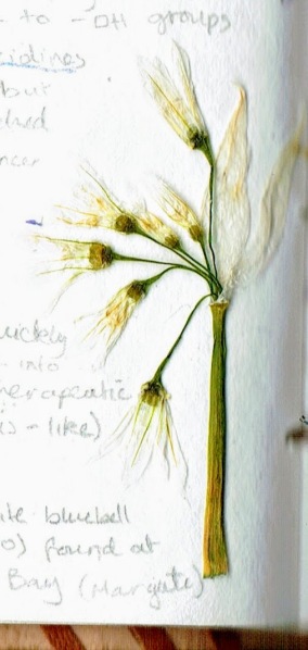
One of the major toxic compounds present in bluebells are polyhydroxylated pyrrolidines (such as nectrisine) which are found in the viscous sap that seeps out of the plant’s nodding stems. These compounds are analogues of sugar but are actually potent glucosidase inhibitors. Due to its many -OH groups, the mammalian body mistakes nectrisine as a sugar and processes it as such – inadvertantly allowing the molecule to interfere with respiration.

In addition to this, polyhydroxylated pyrrolizidines, which have a similar chemical action to nectrisine, are toxic alkaloids that have been proven to poison livestock that have the misfortune of ingesting bluebells. Despite this, these compounds are now being considered for use in cancer therapeutics.

Most fascinatingly, bluebells contain a type of cardiac glycoside, Scillaren A, which can rapidly increase the output force of the heart. Heart cells, myocytes, contain protein pumps embedded in their cell membranes which actively transport sodium ions out of the cell to create an essential electrochemical gradient. In the heart, there is also a sodium – calcium ion exchanger which maintains the ion homeostasis. 

Cardiac glycosides, such as Scillaren A found in bluebells, inhibit the movement of sodium ions out of the cell by effectively disabling the ion pumps. This increases the concentration of sodium ions inside the myocytes which, in turn, increases the calcium ion concentration as sodium ions inside the cell are exchanged for calcium ions.

In myocytes, the accumulation of calcium increases the power with which the cells can contract (contractility) – increasing cardiac muscle contraction which can be crucial in controlling the heart rate in cases of arrhythmia and even chemically restarting the heart in atrial fibrillation.

The cardiac action of Scillaren A is stronger than that of the commonly used digoxin, which is derived from foxglove, and is ideal for situations in which digoxin alone is insufficient or the patient has a digitalis intolerance. Additionally, if an improper dosage is administered, Scillaren A is poisonous but, unlike many other cardiac glucosides, is non-lethal as it is poorly absorbed into the gut. The compound also has a very high therapeutic index as it is rapidly eliminated from the body. All of these qualities make bluebells a fundamentally desirable pharmaceutical source, however, since native bluebells are an endangered and protected species, bluebells cannot be harvested to produce medication.

bee - Edited (1)

Conservation efforts are essential to protect and preserve this fragile, flowering species so that we may be able to enjoy their magnificent appearance and benefit from their therapeutic value in the coming decades.


* White bluebells which lack pigmentation also exist albeit they are somewhat rare with only 1 in every 1000 native bluebells being white. Below is a pressed white bluebell I came across in Margate. bluebell - Edited (2) (1)

Dandelion: Taraxacum Officinale

Standard

[This posts features my original botanical sketches]

For me, the most marvellous manifestations of spring are the bright dandelions emerging from previously barren, frost-glazed soil – bringing promises of brighter days to come.  The humble dandelion is so often maligned as a weed, however these perennial plants have a rich therapeutic history.

dandelion - Edited (2) (1)

Nearly half a millennium ago, Nicolas Culpeper, a prominent herbalist of the early 1600s, noted that the dandelion has “an opening and cleansing quality” and remarked upon its efficacy at treating liver ailments (including cirrhosis) as well as diseases of the spleen and gall-bladder. Many of Culpeper’s contemporaries, and indeed modern herbalists, also acknowledge the powerful diuretic properties of dandelion leaves and root.

Dandelions have even proven valuable in cutting-edge pharmaceuticals. Researchers from the Department of Chemistry and Biochemistry at the University of Windsor, Canada,  have found that dandelion root extract can efficiently kill certain types of aggressive leukaemia cells. This extract can induce apoptosis (programmed cell death) and autophagy (wherein the cancer cell breaks down its own organelles), and even disrupt mitochondrial membranes. The lack of toxins and alkaloids in the plant, combined with the extract’s selectivity for leukaemia cells, makes the dandelion a particularly promising candidate for safe chemotherapy applications.


Ovadje, P., Hamm, C., & Pandey, S. (2012). Efficient induction of extrinsic cell death by dandelion root extract in human chronic myelomonocytic leukemia (CMML) cells. PloS one7(2), e30604. doi:10.1371/journal.pone.0030604

https://www.ncbi.nlm.nih.gov/pmc/articles/PMC3281857/